Plankton Pioneers

A substantial part of being a scientist, especially when you are relatively new, is learning about the research that has been done over the years. The purpose of this is to critically examine the published scientific work and identify the knowledge gaps that still remain. On this blog, we talk a lot about plankton images and all of the interesting data that you are helping us analyze. But what got us started down this path? How did scientists realize that there was a completely different world other plankton samplers (nets) were missing?

With improvements in SCUBA diving gear and underwater photography, the 1970s were an exciting time to be a marine biologist. A handful of young scientists, who would later become some of the leading experts in their field, pioneered the use of “blue water diving.” This involves divers going out to the open ocean, descending to a particular depth, and observing the life around them. Blue water diving is little bit more difficult than your typical dive on a coral reef because there is no reference point for your eye to detect if you are ascending or descending in the water. They had to be very good at controlling their buoyancy!

Dr. Alice Alldrege, now a professor at UC Santa Barbara, counts plankton within a fixed volume to estimate their concentrations

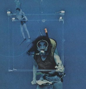
Alice Alldredge, a research professor at UC Santa Barbara, counts plankton within a fixed volume to estimate their concentrations

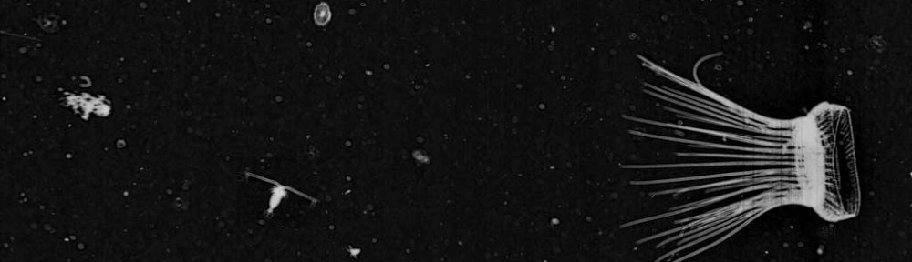
The amazing things the scientists witnessed, and that many of you get to see in the ISIIS images, were documented in an article in National Geographic in 1974, written by Dr. William Hamner. The article effectively captures the sense of wonder and amazement that they experienced observing these fragile animals up close. They departed from Bimini, Bahamas and would dive on the side of the island protected from the strong Gulf Stream current. Each researcher focused on a different plankton group, capturing a variety of specimens and estimating their abundance. During their research, they were the first people to image larvaceans swimming freely in the ocean, as well as discovering abundant pteropods once thought to be very rare. They even occasionally saw curious sharks passing by!

Hamner1974pic2

Salp reproducing a new chain asexually – photo credit LP Madin

Ctenophore ocyropsis maculata with its lobes open.. The scientists discovered that this species uses its lobes primarily for locomotion

Ctenophore Ocyropsis maculata with its lobes open. The scientists discovered that this species uses its lobes primarily for locomotion.

The data gathered by these “Plankton Pioneers” was so valuable that later, when computer technology advanced, their work provided justification for developing imaging systems, such as the Video Plankton Recorder (Davis et al. 1992) and ISIIS, that could collect data at a much faster rate than divers. No doubt that as technology advances even more, there will be plenty of discoveries made about the secret lives of plankton. So if you ever have a chance to be in the ocean, take a minute to ignore the pretty corals and fish to look just a few inches in front of your mask. You just might see a whole world most overlooked for so long.

All photos are from National Geographic. Head to your local library and read the entire article! It is definitely worth the trip!

References:

Davis CS, Gallager SM, Solow AR (1992) Microaggregations of oceanic plankton observed by towed video microscopy. Science 257: 230-232

Hamner, WM (Oct. 1974) Ghosts of the Gulf Stream: Blue-water plankton. National Geographic Magazine 146: 530-545

OSTRICH cruise leg 2 almost done!

We have been zigzagging up and down the Florida Straits for the last two weeks. This photo, from two days ago, shows our ship track (in blue) and drifter track (in red).

RV Walton Smith Tracks

Our first study was the Spatial Study, where we deployed a drifter at the beginning of the day, and then followed it, sampling with ISIIS and MOCNESS in a zig-zag track for the whole day. We started our cruise sampling near Miami and Bimini, Bahamas, and finished up the Spatial Study in the FL Keys.

Photo by Cedric Guigand, taken with a Phantom DJI drone

Research Vessel F.G. Walton Smith during the OSTRICH Cruise, June 2014. Photo by Cedric Guigand, taken with a Phantom DJI drone.


In the second leg of our cruise (which concludes tonight), we have been sampling in the Lagrangian Study, which lasted a total of 4 days. We deployed a drifter in the Lower Keys at the beginning of the study and have been following it continuously, sampling with ISIIS and MOCNESS.

The idea is that by following a drifter, we will be continuously sampling the same body of water. Consider this analogy: you are sampling a fast moving stream. If you stand at a fixed point on land and continuously sample, you sample different parts of the stream because it is constantly flowing in front of you. However, if you build a raft and float down the stream with it, sampling along the way, then you are actually sampling the same part of the stream at different points in time. The latter case is what we are doing. The Gulf Stream / Florida Current is our fast moving “stream” and we are drifting with the stream in a fancy raft, sampling along the way.

R/V F.G. Walton Smith, aerial view of the back deck, with ISIIS on board. Photo by Cedric Guigand

Continue following our updates on the ISIIS Facebook page as we conclude our 18-day cruise this week.

ISIIS in the field: OSTRICH cruise in progress

Hi Plankton Portal!

The Science Team is currently out in the field in the Straits of Florida, on the R/V Walton Smith, sampling with both ISIIS and MOCNESS (Multiple Opening Closing Net and Environmental Sampling System), on an 18-day cruise titled OSTRICH (Observations on Subtropical TRophodynamics of ICHthyoplankton).

OSTRICH logo

The overall goal of this NSF-sponsored project is to quantify the patterns and consequences of the fine-scale to sub-mesoscale distributions of larval fishes, their prey, and their predators near and across a major western boundary current passing through the Straits of Florida. By sampling a series of water masses at very high resolution, this study addresses specific hypotheses concerning: i) the drivers of aggregations and patchiness, and ii) the biological consequences of predator-prey interactions at fine scales.

postdoc ad sample images

Sample ISIIS images showing diversity of plankton from multiple coastal sites (including the Southern California Bight!)

Sampling involves a novel combination of detailed in situ sampling of the horizontal and vertical distributions of plankton, targeted fine-scale net sampling, and analyses of individual-level recent daily larval growth to enable the identification of the biological and physical processes driving fine-scale plankton distributions.

Follow along on the ISIIS facebook page as we periodically post updates (via our terrible internet connection at sea!) and also check out this cool video made by one of our cruise participants, Chris Muiña:


Plankton blooms: Causes and Consequences

As organisms that cannot swim against the currents, plankton are intimately connected to their physical environment. Many species are quite sensitive to the temperature, salinity, and nutrient levels that either lead to their proliferation or demise. Physical conditions and nutrient levels can lead to high abundances of particular plankton types. These plankton “blooms” are common throughout the world’s oceans and can be composed of phytoplankton, zooplankton, or gelatinous zooplankton, depending on the environmental conditions.

Generally phytoplankton (plankton that use photosynthesis like plants) need nutrients and light to grow at very high rates. Since light is readily available in the surface ocean, nutrient availability is the most important driver of phytoplankton blooms. Phytoplankton blooms of most concern to environmental monitoring groups are often described as Harmful Algal Blooms (HABs). Some HABs composed of diatom species Pseudo-nitzschia spp. or the dinoflagellate Karenia brevis can produce toxins harmful to copepods, fish, and higher trophic levels like dolphins and humans. “Red tides” are actually blooms of Karenia brevis that sometimes lead to massive fish dieoffs. Other phytoplankton blooms are harmful not because of the toxins that they produce, but because of the processes that happen when the blooms die off: massive amounts of phytoplankton die and sink to the bottom where they are decomposed by bacteria. These bacteria use oxygen to consume the dead phytoplankton, creating large portions of the water column that are low in oxygen. Fishes and some zooplankton avoid these low oxygen zones, but gelatinous zooplankton seem to be able to withstand low oxygen conditions. These low oxygen regions are often referred to as “dead zones” because very few animals can live there. A dead zone occurs regularly in the summertime in the northern Gulf of Mexico and has been expanding in recent years. Reducing nutrient/fertilizer runoff from farmlands and cities is therefore crucial to limiting the growth of phytoplankton and maintaining healthy coastal ecosystems.

A bloom of Karenia brevis viewed from the air. This "crimson tide" is only composed of small dinoflagellates, but it can have devastating consequences for a coastal ecosystem. Source: http://microbewiki.kenyon.edu/index.php/File:Red_tide_genera.jpeg

A bloom of Karenia brevis viewed from the air. This “crimson tide” is only composed of small dinoflagellates, but it can have devastating consequences for a coastal ecosystem.
Source:
http://microbewiki.kenyon.edu/index.php/File:Red_tide_genera.jpeg

Blooms of zooplankton can form via two different mechanisms 1) currents from different water masses merge to create a dense patch of organisms, or 2) consistently favorable conditions allow the zooplankton to reproduce faster than their predators can consume them. These two mechanisms are distinguished in the scientific literature as “apparent blooms” and “true blooms” (Graham et al. 2001). Apparent blooms can result from converging currents such as fronts, or the behavior of zooplankton aggregating along some kind of physical discontinuity, such as a thermocline. True blooms are typically the result of high food concentrations, high survival of larvae/juveniles, or a combination of multiple factors. For scientists, it is sometimes difficult to know what mechanism led to a zooplankton bloom, and they need to consider the history of the water masses where the zooplankton are found to figure out how the bloom formed (Greer et al. 2013). Zooplankton blooms associated with copepods are generally considered to be healthy for the ecosystem. The timing of copepod peak abundances with the first feeding of larval fishes is thought to be an important factor contributing to the variation in fish population abundances (Cushing 1975). Blooms of jellyfish, on the other hand, are often associated with ecosystems that are environmentally degraded through high nutrient input or consistent overfishing (Jackson et al. 2001), but there are some scientists who think jelly blooms are simply a characteristic of their life histories (Condon et al. 2013). More data on jellyfish abundances over longer time periods will help scientists understand relationships between the environment and the frequency of jellyfish blooms. You can also report jellyfish sightings at http://www.jellywatch.org.

Here is avideo of one ISIIS downcast through the water column of near a frontal feature offshore of San Diego, CA, USA. It shows a bloom of Solmaris spp. jellies concentrated near the surface. Convergent currents that are commonly seen at fronts likely contributed to the formation of this bloom.

References:

Condon RH, Duarte CM, Pitt KA, Robinson KL, Lucas CH, Sutherland KR, Mianzan HW, Bogeberg M, Purcell JE, Decker MB, and others (2013) Recurrent jellyfish blooms are a consequence of global oscillations. Proc Natl Acad Sci U S A 110:1000-1005

Cushing DH (1975) Marine ecology and fisheries. Cambridge University Press, London

Graham WM, Pagès F, Hamner WM (2001) A physical context for gelatinous zooplankton aggregations: A review. Hydrobiologia 451:199-212

Greer AT, Cowen RK, Guigand CM, McManus MA, Sevadjian JC, Timmerman AHV (2013) Relationships between phytoplankton thin layers and the fine-scale vertical distributions of two trophic levels of zooplankton. Journal of Plankton Research 35:939-956

Jackson JBC, Kirby MX, Berger WH, Bjorndal KA, Botsford LW, Bourque BJ, Bradbury RH, Cooke R, Erlandson J, Estes JA, and others (2001) Historical overfishing and the recent collapse of coastal ecosystems. Science 293:629-637

Great plankton pictures taken offshore Miami

Here is a nice collection of plankton underwater shot  by Dr. Evan D’alessandro while conducting research offshore Miami

140304 Fowey Rocks IMG_6911

the Portuguese Man of War is not a Jellyfish but a Siphonophore!

The Portuguese Man of War or Physalia is a siphonophore floating at the surface of the ocean like a drifting balloon with deadly tentacles waiting to capture careless preys. Its body looks like a sail that catches the ocean wind to propel itself.

More Information on Physalia

140304 Fowey Rocks IMG_6779

Portuguese Man of War Fish

The Portuguese Man of War Fish or Nomeid seems to be immune to the powerful sting of the Physalia. It is actually a very agile swimmer that can avoid the stinging tentacles.It uses this deadly siphonophore for shelter against predators and can also feed on some of the smaller tentacles that do not seems to have a strong sting.

140304 Fowey Rocks IMG_6950

Nice ctenophore

Our favorite, the Venus Belt! Nice to see this one in color!

140304 Fowey Rocks IMG_6976

salp chain

If you click on the this amazing picture you will see a small fish larvae seeking shelter among the salps. very neat!

140304 Fowey Rocks IMG_6993

ctenophore bloom

Beautiful bloom of lobate ctenohphores. The wall of death some smaller plankters:)


Arrow worms: voracious plankton predators

You may think orcas or great white sharks are the most voracious predators in the oceans, but based on their abundance and ability to consume a wide range of prey items, chaetognaths (a.k.a. “arrow worms”) give those big animals a run for their money. Large predators like sharks are extremely rare, but scoop up a bucket of seawater almost anywhere in the world and you are likely to find a few chaetognaths (if you have a microscope handy). Chaetognaths are transparent worms that often remain motionless in the water column, apparently relying on the element of surprise to capture a wide variety of plankton, including copepods, appendicularians, small fish larvae, and smaller chaetognaths. Chaetognaths are thought to be generalist feeders because their stomach contents often reflect the community captured by plankton nets. They use a mass of chitinous hooks around their mouths to capture prey – which gives them their name (“chaetognath” translates from Latin to mean “hairy jaw”) and a notoriously menacing appearance.

Chaetognaths are often straight in the ISIIS images but can also swim rapidly for short distances. The camera typically cannot resolve the tiny chitinous hooks on the chaetognath's mouth.

Chaetognaths are often straight in the ISIIS images but can also swim rapidly for short distances. The camera typically cannot resolve the tiny chitinous hooks on the chaetognath’s mouth.

Chaetognaths comprise about 100 species that are all typically 1-2 cm long. They are most abundant along the coasts, with some species being so sensitive to salinity that oceanographers can identify discrete water masses based solely on the community of chaetognath species. Similar to many other types of zooplankton, chaetognaths are hermaphrodites, first being male then changing into female as they get larger. Fertilized eggs can be attached to vegetation or encased in a gelatinous web. Eggs then hatch into juvenile chaetognaths, and thus they have no larval stage. This is called direct development because there is no process of metamorphosis.

A clear image of the chaetognath's mouth on the cover of Current Biology.

A clear image of the chaetognath’s mouth on the cover of Current Biology.

The chaetognath’s body is streamlined and adapted to feeding with minimal visual input. The have sensory cilia that can detect small vibrations in the water that tell the chaetognaths that prey is within striking distance. With a quick flick of its tail, the chaetognath surges forward to capture the prey in its chitinous hooks used for grasping. It then transfers the prey to its mouth where it is swallowed whole. Some deeper water chaetognaths (>700 m deep) can even use bioluminescence to create a cloud of light that scientists think can be used to escape predation (Haddock and Case 1994).

The most handsome chaetognath found by our citizen scientists!

The most handsome chaetognath found by our citizen scientists!

References:

Haddock SHD and Case JF (1994) A bioluminescent chaetognath. Nature 367:225

Johnson WS and Allen DM (2005) Zooplankton of the Atlantic and Gulf coasts: A guide to the identification and ecology. Johns Hopkins University Press, Baltimore, MD

Lalli CM and Parsons TR (1997) Biological oceanography an introduction. Elsevier Butterworth-Heinemann, Burlington, MA

Plankton Portal “en Français” (in french) coming soon!

plankton french

Artwork by Jean-Olivier Irisson

Great news! We are working on translating Plankton Portal in French with our
French Collaborators: Fabrice Not from the Station Biologique de Roscoff and Jean-Olivier Irisson from the Observatoire Océanologique and Station Zoologique de Villefranche-sur-mer. “The idea is also to get some interest from French schools to develop a curriculum around Plankton Portal” Dr. Irisson explains.  Stay Tuned.