Shedding light on the diverse plankton universe of the Northern California Current

Moritz S Schmid* and Margaret E Martinez

* schmidm@oregonstate.edu

The northern California Current (NCC) is a dynamic, highly productive region within the broader California Current Large Marine Ecosystem (CCLME), that exhibits strong ecosystem variability on seasonal, interannual, and decadal time scales. Along the California, Oregon, and Washington coasts, wind blowing alongshore the US coast can create upwelling and downwelling events. Wind blowing Southward pushes surface water away at a 90-degree angle (through the Coriolis force, see more info here). Surface waters moving away are then replaced by the deeper water underneath. This deeper water coming to the surface has more nutrients than the surface water (because the nutrient-using phytoplankton doesn’t grow as good at depth – without sunlight), and thus this upwelled, nutrient-rich water can now sustain new phytoplankton growth, which in turn can then support more zooplankton growth, and so on. Downwelling on the other hand occurs when the alongshore wind is blowing Northward. The same Coriolis effect now pushes water into the landmass at a 90-degree angle from the Northward blowing wind. Because the water has nowhere to go, the least resistance for the water is to push downward. Thus, surface water is forced deeper, and no upwelling effect occurs. Along the CA, OR, and WA coasts, wind patterns differ, and while most of the California coast experiences Southward blowing winds year-round, and subsequent upwelling also occurring year-round, Oregon and Washington experience a mix of North-, and Southward winds in summer, and predominantly Northward winds winter. The mix of South-, and Northward wind means the coast gets some upwelling and some downwelling, also referred to as intermittent upwelling, while Northward winds in winter mean that the coast gets less new nutrients from depth during that time. Ultimately, this means that the California coast experiences continuous new nutrient input from deep water that can be used by the organisms, while along the OR and WA coasts this input is more inconsistent. Nonetheless, also OR and WA coastal waters provide a unique and productive environment for plankton and fishes that sustains the livelihood of many communities on the Pacific Northwest coast.

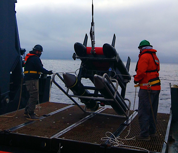
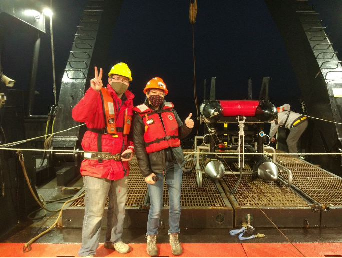
In May and early June 2021, we joined the National Oceanic and Atmospheric Administration’s (NOAA) Northern California Current (NCC) cruise aboard NOAAS Bell M. Shimada. NOAA’s NCC cruises occur on a regular basis and are designed to characterize the planktonic ecosystem ranging from northern California to Washington, with a focus on the Newport Hydrographic Line. Our aim was to collect zooplankton imagery for the NCC Marine Biodiversity Observation Network (MBON, see more info here and here) funded by NASA, as well as the Belmont Forum-funded project ‘World Wide Web of Plankton Image Curation’ (wPIC, see more info here and here), using the In Situ Ichthyoplankton Imaging System (ISIIS, Figs. 1-4).

The MBON project’s focus is on seascapes. Similar to different landscapes on land, the surface ocean can be classified into different categories based on environmental variables such as phytoplankton abundance and temperature. While seascapes are currently classified using data from the surface ocean (i.e., the first 10 m of the ocean) largely due to heavily relying on satellite data, we collect data using ISIIS to inform seascapes also using information from deeper waters (e.g., data on plankton and temperature down to 100 m). wPIC is aiming at very different things – it is largely a project focused on underwater imaging methods. The idea is that all around the world laboratories use different instruments for imaging aquatic animals, and these instruments also come with different methodologies of processing the images and ancillary data. wPIC’s aim is to streamline these efforts more and to better exploit synergies between different projects and instruments. wPIC includes project partners from France, Japan, Brazil, and the US.

Figure 1. Deploying ISIIS on a gray day off the Oregon Coast.
Figure 2. ISIIS is secured to the aft deck of NOAAS Bell M. Shimada as it heads out past the bar in Newport, Oregon, for the start of the Spring 2021 NCC cruise.
Figure 3. Moritz and Margaret posing with ISIIS after a successful nighttime recovery.
Figure 4. “Flying” ISIIS means observing multiple monitors, each tracking several variables, ranging from ship and ISIIS speeds, communication with the winch operator outside who hauls-in and pays-out oceanographic wire, raising and lowering the ISIIS, to making sure all cameras are recording, and looking for interesting ecological features in the live imagery coming through the fiber optic data stream.

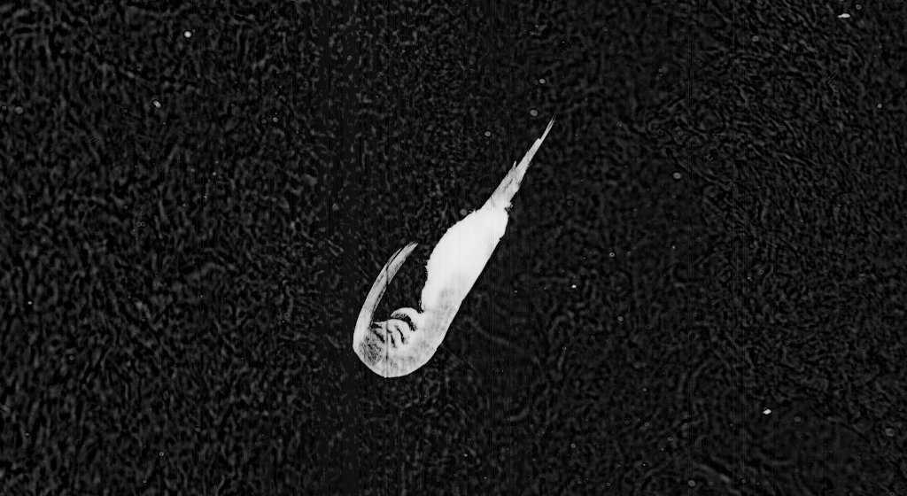
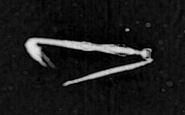
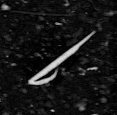
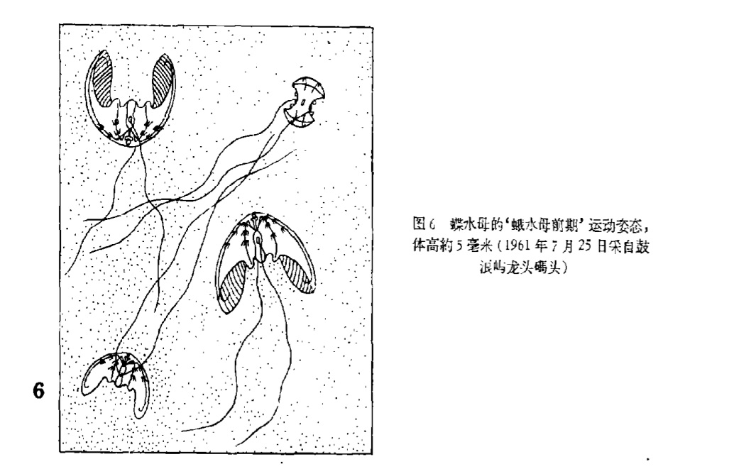
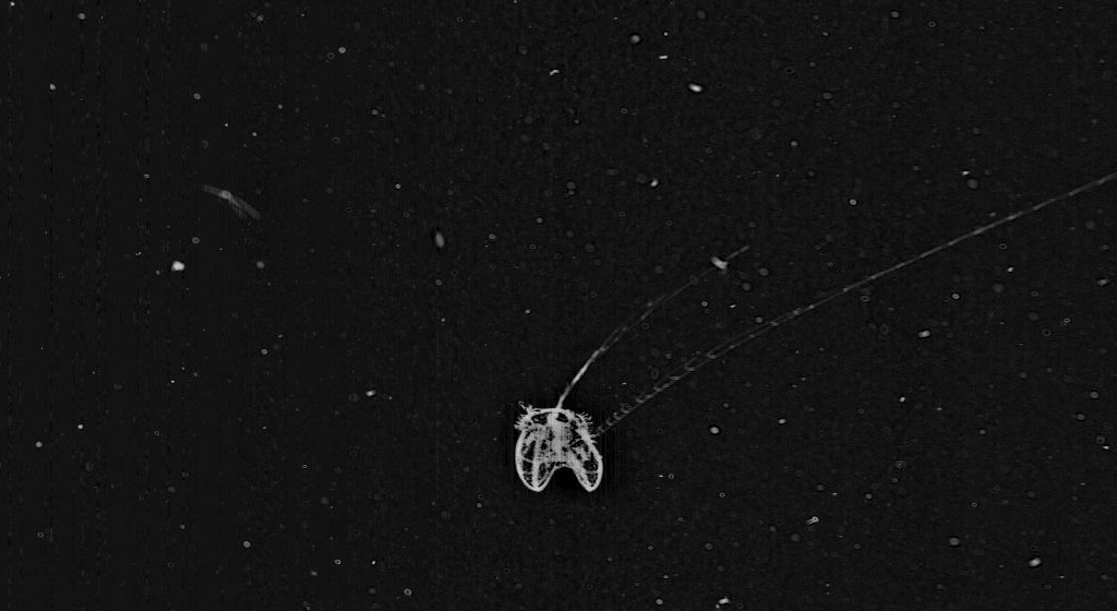
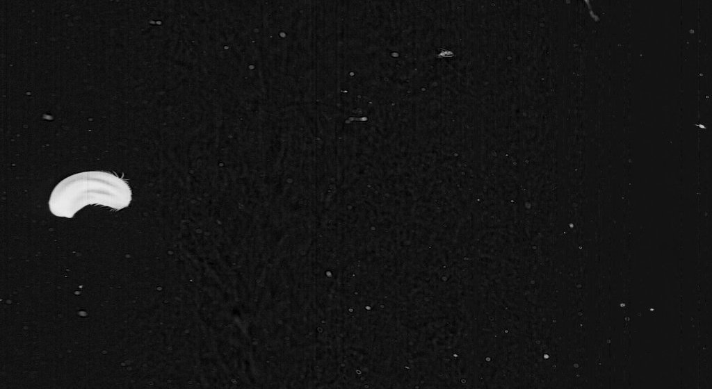
ISIIS is a line-scan and shadowgraph imager that records the shadows of organisms swimming through the beam of light projected by a LED (Fig. 5). With this setup, a water parcel measuring 13 cm x 13 cm x 45 cm is recorded by the camera system, ultimately leading to 180 L of seawater imaged per second. Compared to other imaging systems, this is a very large volume of imaged water, which gives us the ability to look at rather rare larval fishes (Fig. 6) as well as fragile jellies and the ubiquitous crustacean zooplankton (Figs 7-8).
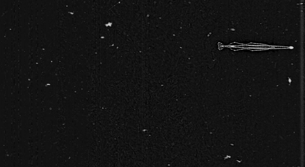
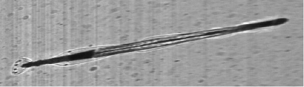
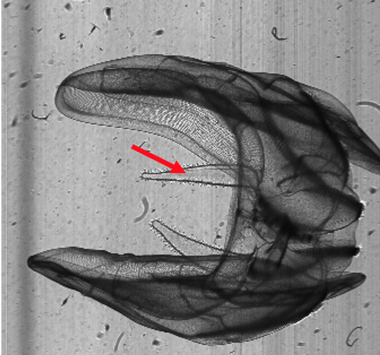
Figure 5. A schematic of the lower pods of ISIIS (compare with Fig. 1) housing the LED and camera setup.

Figure 6. A myctophid (lanternfish, left), and an engrauliid (anchovy, right).
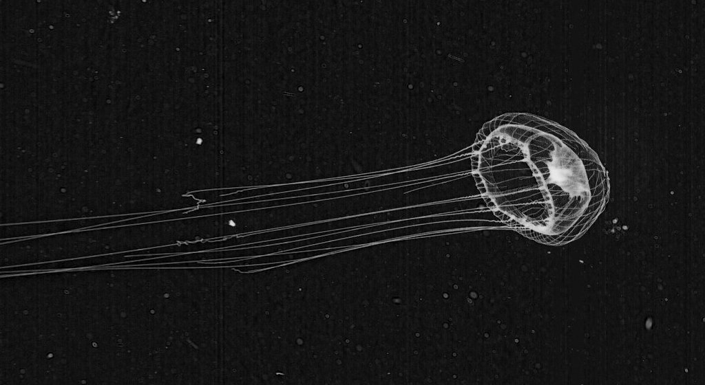
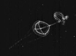
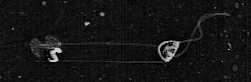
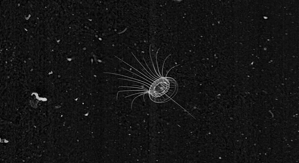
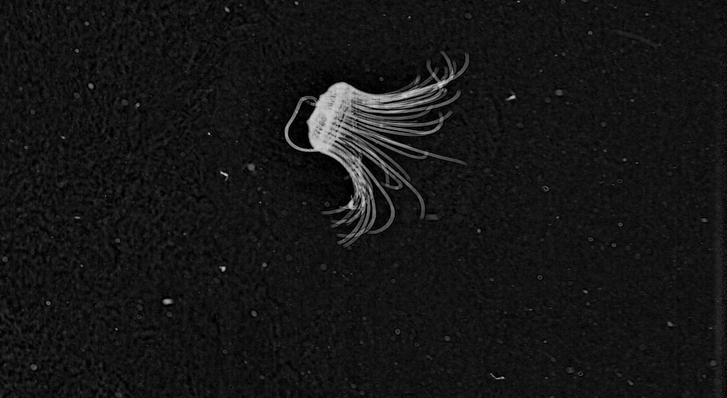
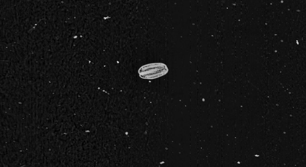
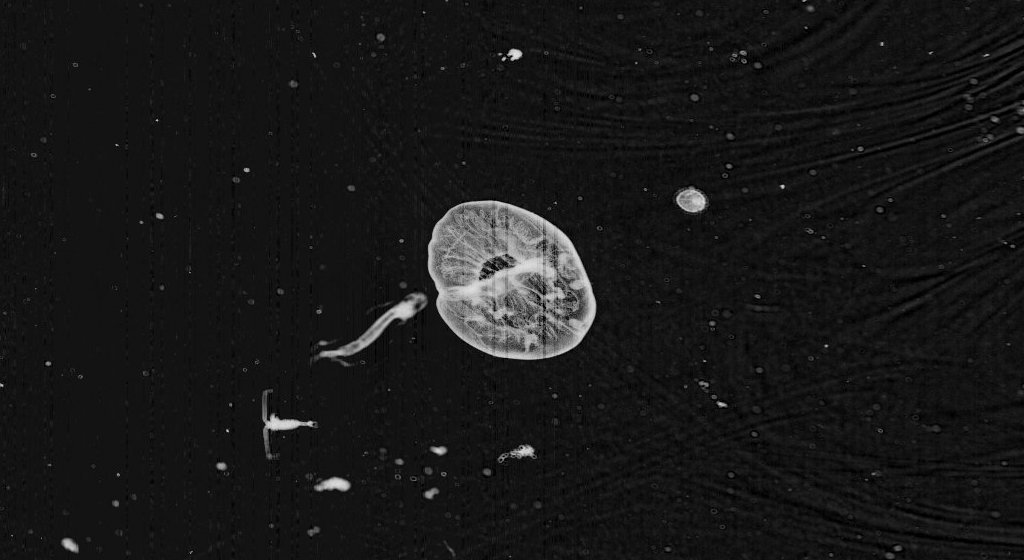
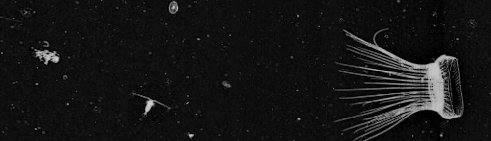
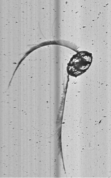
_________________________________________________________________________________

A dense layer of doliolids.
A chaetognath (arrow worm).
A cydippid ctenophore.
Eutonina indicans, a leptomedusae, with gonads visible in the radial canals.

_________________________________________________________________________________

Figure 7. Key gelatinous zooplankton in the NCC includes doliolids, chaetognaths, ctenophores, and hydromedusae*.
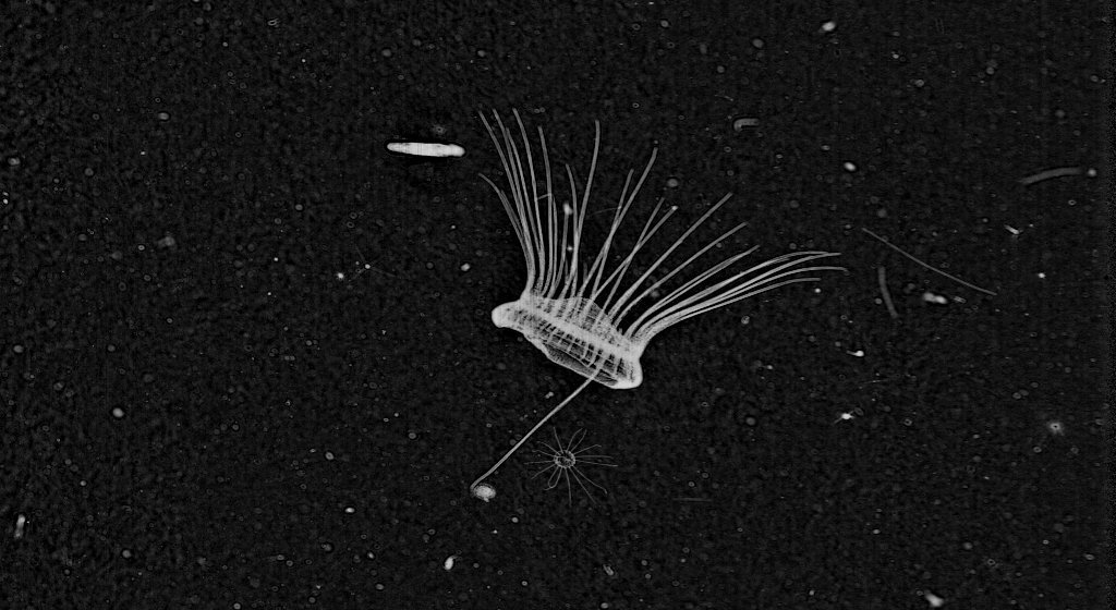
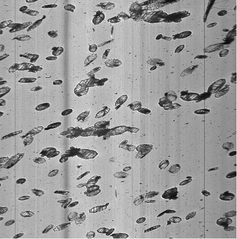
_________________________________________________________________________________

Several euphausiids having a dance party.
Mitrocoma cellularia.

A large euphausiid dwarfs a calanoid copepod.
A lobate ctenophore showing off its auricles (red arrow).

_________________________________________________________________________________

Figure 8. Key crustaceans include euphausiids and calanoid copepods. ISIIS’s ability to image fragile jellyfish without disturbance is clearly visible in the images of Mitrocoma and its many tentacles, as well as a ctenophore with its auricles clearly visible*.

*All images except the doliolids (Crescent City line, 5/26/2021), were taken from Newport Hydrographic Line imagery (5/23/2021).

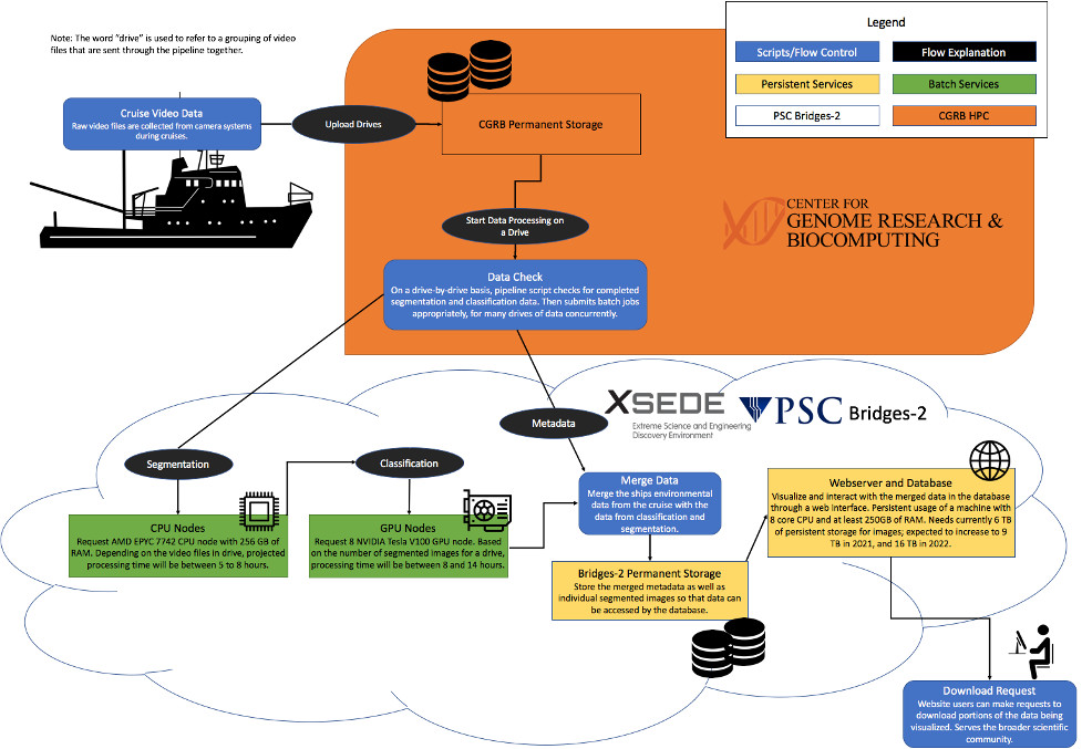
Using a sophisticated image processing pipeline (Luo et al. 2018, Schmid et al. 2021), based on artificial intelligence, we can quickly identify all plankton in the images, and also get their sizes. These ecological data can then be used in a diversity of ecological studies. As ISIIS produces vast quantities of data (ca. 75-100 million images for a 7-hr ISIIS transect) we collaborate with the National Science Foundation’s Extreme Science and Engineering Discovery Environment (XSEDE), a nationwide supercomputing cluster. Controlling the processing from Oregon State University’s (OSU) Center for Genomics and Biocomputing (CGRB), but utilizing hardware at XSEDE (Fig. 9), we are able to get through the data blazingly fast. The resulting data enables us to tackle ecological questions that wouldn’t be possible with more traditional net systems (e.g., Briseño-Avena et al. 2020, Schmid et al. 2020, Swieca et al. 2020).

Figure 9. The Plankton Ecology Lab’s image processing pipeline deployed at OSU’s CGRB and using resources on NSF XSEDE. The orange panel shows components at the CGRB, while components in the lower panel (i.e., the cloud) are deployed on NSF’s XSEDE supercomputing cluster.

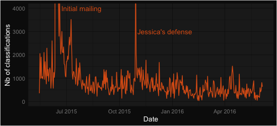
At this point we want to give a big shout out to all our citizen scientists at Plankton Portal who help with making these studies possible by volunteering to classify plankton. This effort helps us to fine-tune plankton classification image libraries and models, and provides insight into how automated plankton classifications differ from human annotations (Robinson et al. 2017). We hope that you enjoyed this little glimpse into collecting the images that you get to look at. We also thank Jennifer Fisher, the Chief Scientist on NOAA’s NCC cruise on NOAAS Bell M. Shimada, as well as Bell M. Shimada’s CO Amanda Goeller, and crew, as well as all scientists who helped with manning the ISIIS winch.

References:

Briseño-Avena C, Schmid M, Swieca K, Sponaugle S, Brodeur R, Cowen RK. 2020. Three dimensional cross-shelf zooplankton distributions off the central Oregon coast during anomalous oceanographic conditions. Progr Oceanogr 188:102436 https://doi.org/10.1016/j.pocean.2020.102436

Luo JY, Irisson J-O, Graham B, Guigand C, Sarafraz A, Mader C, Cowen RK. 2018. Automated plankton image analysis using convolutional neural networks. Limnol Oceanogr Methods 16: 814-827 https://doi.org/10.1002/lom3.10285

Robinson KL, Luo JY, Sponaugle S, Guigand C, Cowen RK. 2017. A tale of two crowds: Public engagement in plankton classification. Frontiers Mar Sci 4:82 https://doi.org/10.3389/fmars.2017.00082

Swieca K, Sponaugle S, Briseño-Avena C, Schmid M, Brodeur R, Cowen RK. 2020. Changing with the tides: fine-scale larval fish prey availability and predation pressure near a tidally-modulated river plume. Mar Ecol Progr Ser 650:217-238 https://doi.org/10.3354/meps13367

Schmid MS, Daprano D, Jacobson KM, Sullivan CM, Briseño-Avena C, Luo JY, Cowen RK. 2021. A Convolutional Neural Network based high-throughput image classification pipeline – code and documentation to process plankton underwater imagery using local HPC infrastructure and NSF’s XSEDE. [Software]. Zenodo. http://dx.doi.org/10.5281/zenodo.4641158

Schmid MS, Cowen RK, Robinson KL, Luo JL, Briseño-Avena C, Sponaugle S. 2020. Prey and predator overlap at the edge of a mesoscale eddy: fine-scale, in-situ distributions to inform our understanding of oceanographic processes. Sci Rep 10:921 https://doi.org/10.1038/s41598-020-57879-x

Happy anniversary Plankton Portal 2.0!

Blog post by Jean-Olivier Irisson

A year ago, we announced Plankton Portal 2.0, which featured a more streamlined design, a simpler tagging interface, and most importantly, a whole new dataset. Since then, this new data from the Mediterranean Sea has spurred a lot of interest and plenty of new questions. Participants on the site were surprised by the difference in size of everyone’s favourite jellies, the Solmarisidae (Solmaris rhodoloma in California, Solmissus albescens in the Med), which are much larger! Siphonophores also seem more abundant there. And the Mediterraean data came with brand new categories of organisms to mark: nice and cute medusa ephyrae (i.e. baby jellies), elegant Pteropods and the elusive fish larvae.
In total, as of last Sunday, 368,361 organisms were marked, on 50,519 distinct images. Through time, the classifications were marked by two peaks in activity: a huge one when the new version was announced through a mailing to the Zooniverse community (thanks everyone!) and another one when we pushed for 1,000,000 classifications in total, to celebrate Jessica’s PhD defense. When we zoom in, we see activity fluctuating around 1000 and now 500 classifications per day. This is still great (but coming back to 1000 would be even better! 😉 ).
time_series
time_series_zoomed


The top 11 contributors, all authors of over 5000 classifications each, are displayed below. If you made this top 11, we owe you special thanks (and probably a beer too). We hope you will stay interested and involved in this project. If you did not, you should really not be disappointed because all other volunteers still collectively account for 60% of the classifications; so you matter very much! Hopefully all of you will be happy to see some of the outcome of your work below.
who


Time for a bit of science! The most common classification was… nothing, empty, zero, zlich, zip… Well, you get the idea. Indeed, when we film the sea, we most often see nothing (nothing living at least). Even though we pre-selected potentially interesting frames for Plankton Portal (the ones having some kind of large object in them), about a third of your classifications did not contain any organism we were interested in. In real life, the proportion of dead detritus vs. living organisms is more around 95% vs. 5%, so our pre-filtering still avoided you a lot of blank frames! In terms of organisms, the 10 most abundant are shown in the figure below.

what


Doliolids, Copepods, and Radiolarian colonies dominate the rest. We immediately noticed, when we shot the images, that Doliolids were particularly abundant. Those organisms are very effective filtering machines and they may therefore have an impact on the density of smaller organisms, in particular unicellular algae. The relative abundance of Copepods and Radiolarian colonies is to be interpreted carefully: Radiolarian colonies can be large and span several frames (therefore increasing the total count) and Copepods are likely under-estimated because we mostly see the larger ones with ISIIS, and they are not the dominant ones in the Mediterranean. Still, it echoes nicely a recent Nature paper by Tristan Biard (a contributor to PlanktonPortal’s talk, under the username Collodaria), which showed that Rhizaria (a large taxonomic group to which Radiolarians belong) can be equivalent in biomass to Copepods, who were previously thought to largely dominate the plankton. These findings were also based on in situ images, because these fragile Rhizaria cannot be collected with nets.


Finally, the images in the Mediterranean were collected along transects (i.e. straight lines) perpendicular to the shore. We were interested in how organisms were distributed along a gradient between coastal and open ocean conditions. In the plots below, the coast is on the left, the open ocean on the right and the vertical direction is depth (top: surface; bottom: 100 m depth). So you basically see a “slice” of water along which ISIIS undulated. The size of the dots is proportional to the number of classifications recorded. You can immediately notice that Doliolids (first plot) are concentrated near the surface, and fish larvae (second plot) even more so! This is a surprising finding for fish larvae, which sometimes ended up in concentrations of over 10  individuals per cubic meter, a number much higher than what was previously observed elsewhere, with conventional plankton nets.
distrib_doliolid_w_icondistrib_fish_w_icon
Radiolarian colonies, on the opposite, tend to be concentrated in mid water (see figure below). Within this messy picture, some structure seems to emerge. Indeed, the white lines on top of the plot are contours of the concentration of Chlorophyll A in the water (i.e. of the amount of unicellular algae). If you look carefully, you will see that those lines are moving up, towards the surface, as we travel offshore (from left to right on the plot). This is actually well known in this region. What is interesting is that the radiolarians seem so follow the same pattern, and that higher concentrations of colonies sit on top of this high Chlorophyll region. Something is definitely going on between these two!
distrib_radiolarian_colonies_w_icon
That’s it for now — thanks again to everyone for this wonderful year of activity! We apologise for not being as active as we would like to be on Talk. To that end, we thank the active moderators who take over this important responsibility. And finally, we thank Zooniverse for the great opportunity and community they created. Now, on to next year!

Fantastic Find Fridays: Feb 2016

Hey plankton hunters!  We are bringing you another round of Fantastic Finds from the Plankton Portal.  Citizen scientists continue to reel in new captures of some truly awesome plankton.  Here are just a few neat finds, ID’s, and novel taxa:

Pteropod mollusk

pteropod

Did you ever learn about marine butterflies in grade-school?  Well good, because there is no such thing as a marine butterfly.  This elegant-looking critter is a pteropod, a type of gastropod mollusk—in other words, a slug!  These mollusks are highly adapted for life in the water-column, as you can see from the butterfly-like wings, (or “parapodia” to a malacologist).  The pteropod wings are actually a highly-modified molluskan foot, i.e. the muscular and slime-secreting mass that slugs glide on.  Evolution really did these slugs a favor, as I do not think anyone could say “ewww!” to such a beautiful animal.

Calycophoran Siphonophore

caly_sipho

Now this is a fantastic image.  A close-up, finely-detailed capture of the head (nectosome) portion of a calycophoran siphonophore—so aptly referred to as a “rocket-ship sipho” here on the Plankton Portal.  The two siphon-like features propelling this colonial critter are very apparent in this image.  Maybe, in truth, siphon-ophore is a pretty apt name for this plankter as well.

Ctenophore: Thalassocalyce inconstans

thalass

Thalassocalyce inconstans is a predatory species of ctenophore, captured feeding in this frame.  The body of the ctenophore is contracted and engorged about the anteroposterior (vertical) axis, giving it the appearance of an inflated, heart-shaped balloon.  Within the fragile and transparent body, you can see the 8 condensed comb rows captured as an array of ragged segments crowning the aboral end.  Fine mesenterial canals also appear as contoured markings that line the engorged body. Ctenophores are tactile predators, meaning all predatory behavior is triggered by physical, non-visual stimulus.  Something in the water column bumped into this Thalasso and got it all riled up, providing ISIIS a great opportunity for this detailed capture of foraging behavior.  If we had a hydrophone for this deployment, I am fairly certain a satisfied lip-smacking would be recorded in a few seconds.

Copepods: Families Eucalanidae and Metridinidae. 

copepods

Copepods are abundant in these ISIIS data, and it is easy to forget what a broad diversity of these important crustaceans are classified on the site.  Here we have two broadly identifiable PP copepods for sample.  The image on the left shows a copepod belonging to the family Eucalinidae: it has a narrow, torpedo-shaped body and the anterior end of the head forms a pointed-triangle.  We think this critter might be a Rhincalanus spp.—if you look close you might be able to make out a small rostrum-like appendage extending forward and tucked down from the head, as well as what may be lateral spination at the end of each mid-body segment (prosome).  This guy’s cruising, antennas spread out and scanning the surroundings.  On the right, we have a copepod belonging to the family Metridinidae, perfectly poised for the ISIIS cam.  How do we guess this ID?  At the end of this copepods lengthy tail (urosome), look closely at the paired fin-like feature (ferka).   Along the outer edge, right before the separation of the individual ferka, can you make out a small, skirt-like protrusion?  If so, just tell your buddies: “hmm, check out that lengthy urosome and ferka segmentation; it must be a Metrinidae species,” and blamo—you are a crustacean taxonomist!

Anthomedusae: Leukartia spp.

anthomed

At first glance, you might be thinking “is this medusae sticking its tongue out at me?” Or maybe it is sporting a ten-gallon hat?  While I couldn’t blame you for such outlandish assertions (I mean, who would write such silly things?), this odd anthomedusae is readily identified to genus by the conical appendage extending from the bell (“apical process”) and causing much confusion on the Plankton Portal.  In this image we get a great view of many internal and external features of this Leukartia sp., including a crenulated (ragged) bell margin, a tall mouth (“manubrium”) in the center of the bell, and many long tentacles projected both downwards and in front of the bell.

Larvacean and mucous house

larvacean

We find a good deal of larvaceans on the site, but this capture is a real beauty.  Larvaceans are gelatinous plankton that filter-feed on detritus in the water column.  You see the critter poking its head out, like the cap on a rolled-up toothpaste tube?  That’s the larvacean, curled up in preparation to pump surrounding detritus through its elaborate mesh-like mucous house.  For a critter that takes up residency in its own secretion, this guy is pretty adorable!

Physonect Siphonophore

sipho_forage

Now this is quite the fantastic find!  Here we are looking at a large siphonophore projecting numerous tentacles across the frame.  It is all-hands-on-deck for this colonial jelly, as it is putting on a mighty foraging display for us.  The big guy is hungry—watch out, ISIIS.

There have been way too many great images to fit in this small serving of photogenic plankton.  We look forward to serving up more fantastic finds in the future.  Keep exploring, plankton hunters!

ISIIS in the field: OSTRICH cruise in progress

Hi Plankton Portal!

The Science Team is currently out in the field in the Straits of Florida, on the R/V Walton Smith, sampling with both ISIIS and MOCNESS (Multiple Opening Closing Net and Environmental Sampling System), on an 18-day cruise titled OSTRICH (Observations on Subtropical TRophodynamics of ICHthyoplankton).

OSTRICH logo

The overall goal of this NSF-sponsored project is to quantify the patterns and consequences of the fine-scale to sub-mesoscale distributions of larval fishes, their prey, and their predators near and across a major western boundary current passing through the Straits of Florida. By sampling a series of water masses at very high resolution, this study addresses specific hypotheses concerning: i) the drivers of aggregations and patchiness, and ii) the biological consequences of predator-prey interactions at fine scales.

postdoc ad sample images

Sample ISIIS images showing diversity of plankton from multiple coastal sites (including the Southern California Bight!)

Sampling involves a novel combination of detailed in situ sampling of the horizontal and vertical distributions of plankton, targeted fine-scale net sampling, and analyses of individual-level recent daily larval growth to enable the identification of the biological and physical processes driving fine-scale plankton distributions.

Follow along on the ISIIS facebook page as we periodically post updates (via our terrible internet connection at sea!) and also check out this cool video made by one of our cruise participants, Chris Muiña:


Fantastic Finds Friday: February 2014

Hey plankton hunters!  This week we are showing off four exceptional zooplankton found by you, our keen-eyed and inquisitive citizen scientists.  We are amazed at how many plankton species have been uncovered on the site and just how capable you all have been at discerning some truly tricky taxa from the varying forms and shapes captured by the ISIIS camera.  We thank all of the citizen scientists for your participation on the Plankton Portal!  These images found by our citizen scientists continue to excite and we are eager to discover what resides in the thousands of images yet to be seen by human eyes!

Annatiara affinis; Anthomedusa — #Medusa #morethanfourtentacles

51d1bee53ae74008a405aea3

This capture of an anthomedusa is definitely a prime example of how the images captured by ISIIS can be equal measures fine-resolution biological data and one-of-a-kind organismal artwork.  This gelatinous organism is baring all for us in this frame and we get a clear view of not only the striations on the exterior of the bell (the exumbrella) unique to this species and the fully extended tentacles, but also the central gastric pouch (stomach) appearing as the dark mass within the bell and the internal network of radial canals where digested food is transported. I think I can also see this critter blushing as ISIIS takes the snapshot!  This medusa shown here is relatively uncommon in the images provided for you from the Southern California Bight, and we couldn’t be happier that our fantastic and dedicated group of citizen scientists spotted this gelatinous beauty.  Annatiara affinis is a hydromedusa like many of the #4tentacles and #morethanfourtentacle medusae found on the site.  The unique (and photogenic) lines appearing along the exterior of the bell were very helpful in pinning down an ID for this critter.  From what we have seen, this seems to be a rare image captured of Annatiara where the tentacles are fully extended from the margin of the bell, and we are extremely grateful that this lovely jelly was so at ease in front of the ISIIS cam.

Shrimp — #Shrimp

51d1bef33ae74008a4061218

This is one of the largest shrimps I have seen on the portal and provides a great side-view of the crustacean anatomy.  The orientation of this shrimp with the abdomen tucked under the carapace (upper shell) and the antenna trailing sharply away from the head indicates that it is moving rapidly towards its posterior (bottom left of image), using a swimming stroke known as the “cardioid escape reaction”—slapping the abdomen shut and quickly propelling the crustacean away from the perceived danger.  This specific behavior played an important role in the field of neuroscience, in fact.  When it was discovered, this behavioral response was the first example of a “command neuron mediated behavior”— meaning a specific behavioral pattern resulting from the stimulation of a single neuron.  I wonder what stimulated this crustacean’s command neuron? Perhaps it is camera shy.

Arrow Worm / Chaetognath — #ArrowWorm

51d1beff3ae74008a4066981

I’m curious if that dark blob may be some out-of-luck plankter soon to be nabbed by this voracious predator.   I am especially fond of these in focus captures of chaetognaths.  The dart-shaped body and the hydrodynamic taper of the paired lateral fins really show off the sleek and elegant body plan of these brutal invertebrate carnivores.  The chaetognath body has a protective outer covering known as a cuticle, a tough but flexible non-mineral layer exterior to the epidermis.  Chaetognaths are notoriously efficient predators and hunt other planktonic organisms using hooked grasping spines that flank the mouth.  A hood arising from the neck region can be drawn over or away from the hunting spines, much like the action of sheathing and unsheathing a blade.  Equipped with an armor of cuticle and sword-like spines these guys are definitely well suited for combat!

Physonect Siphonophore — #Sipho #Corncob

51d1bdbc3ae74008a401fa38

The siphonophores love to put on a good show for us here on the portal and this frame is truly exceptional.  The shadowgraph imaging technique used by ISIIS lends itself to capturing in detail the elaborate gelatinous structures displayed by these colonial organisms.  Siphonophores are comprised of many single animals, or zooids, which are highly specialized and coordinated in function.  The zooids of a physonect siphonophore arise from a long stem at the end of which is a gas-filled float referred to as a pneumatophore.  The pneumatophore is on display in this image here appearing as the dark, oval-shaped appendage on the upper left end of the main “body.”  The portion that resembles a corn on the cob is referred to as the nectosome.  The nectosome is composed of many swimming bells, or nectophores, each one of which is a single medusoid zooid.  These nectophores display remarkable coordination among each other and the selective contraction of these zooids allows for the siphonophore to move and turn in any and all directions.  Physonect siphonophores are predators and rely on long, branching tentacles for prey capture.  The one whipping across the frame here is definitely on the prowl.  Each tentacle arises from a single feeding polyp situated below the nectosome in a region called the siphosome.  You can see the siphosomal region on this specimen as the narrowing, darkly filled feature curling upward from the base of the nectosome.  They sure have a lot of ‘somes’ and ‘phores’ but we forgive their repetitive nomenclature because we are always glad to find some siphonophores.

We hope this has been a fun and informative look at a few of the many tremendous critters captured by ISIIS and found by the citizen scientists.  If you come across an image you think is particularly cool on the portal then tag it with #FFF and we will check it out for use on the blog.  As always, looking forward to the next Fantastic Find Fridays!

FFF special behavior

Hello everyone. We have a special “behavior” Fantastic Finds Friday (FFF) today! These frames were selected from your posts to illustrate the power of the human eye to detect rare and unusual phenomena. The frames selected here may not be the most beautiful you have seen so far, but the story behind them is fascinating and could not be told without the help of our citizen scientists.

Here is great shot of a larvacean (also known as an appendicularian) getting spooked by the movement of ISIIS. Larvaceans are known to escape from their mucous house if threatened by a predator. Unfortunately the house can’t be used again, and they will start secreting a new house once the threat is passed.

51d1bd9b3ae74008a400dcb1

Arrow worms (chaetognath) are voracious predators capable of engulfing prey as big as their own body. In these images, you can see an arrow worm catching a larvacean and the other grasping what appears to be a copepod. Their mouths resemble a crown of spikes ready to impale any unlucky prey. Chaetognaths also prey on fish larvae.

51d1bd993ae74008a400c7d0 51d1bda43ae74008a4012997

These two medusae just snagged a larvacean house. Accident or deliberate attempt to feed on these poor guys? The long trailing tentacles act like a sticky fishing net that retracts to bring in the catch of the day.

51d1bd993ae74008a400c49e 51d1bed43ae74008a40537f6

These Solmaris seem to be reaching for something (one tentacle pointed opposite to the others). Solmaris have been seen feeding on other jellies – even large siphonophores! They swim with their tentacles forward to maximize the chances of catching a prey. they then move the item to their mouth with one tentacle (like an arm almost).

51d1bed43ae74008a4053599 51d1beea3ae74008a405d135 51d1befc3ae74008a40653ad

No, these are really two different frames! Amazing consistency in posture isn’t it? And look at these two tentacles reaching out – sensing their environment? Hoping to encounter a tasty prey item? If we detect enough of these organisms, we could try to investigate at which time or location they behave this way. This could be a really interesting project!

51d1bd853ae74008a400264a 51d1bd903ae74008a4007734

So if you see something interesting like these example or suspect some interaction is at play in one of the frame use the hashtag #behavior. Remember to mark frames you want considered for future Fantastic Finds Friday posts with #FFF. Thanks, and keep up the good work!

Fantastic Finds Friday #FFF – Cydippid edition

We are nearing the end of Friday, so apologies that this post is late! Hopefully it will be enjoyable for you weekend warriors! By the way, did you see that we are almost at 200,000 classifications?! I am so impressed by this amazing group of citizen scientists that make Zooniverse projects a success, particularly this one. THANK YOU.

We are going to use FFF to point out some amazing pictures that you guys have identified and called to our attention in the last week+, and also to clarify some confusion on a tricky category.

51d1bd833ae74008a40013bd
http://talk.planktonportal.org/#/subjects/APK00003ui

Cydippid ctenophore – #cydippid 

This is a type of comb jelly, called a cydippid ctenophore. We think that this organism is Hormiphora californiensis or a relative. It has a egg shaped body with two tentacles, which are typically extended (for feeding), but also can be retracted into the sides of its body.

The relative of Hormophora californiensis is Pleurobrachia bachei, the sea gooseberry. Check out the following video of P. bachei feeding on some brine shrimp:

Here is another video of P. bachei from the Vancouver Aquarium:

For every easily classified cydippid ctenophore there is also other cydippids that are more difficult to classify by users. These ctenophores include Mertensia and Haeckelia beehleri, which are also cydippids but have their tentacles withdrawn. See below:

51d1bd993ae74008a400ca6a
http://talk.planktonportal.org/#/subjects/APK0000yb7

This is also a cydippid ctenophore – but it has its tentacles withdrawn.

To add some more complication to the matter, there are also some lobate ctenophores, like the one below, whose young have a cydippid-like phase.

51d1bd903ae74008a4007782
http://talk.planktonportal.org/#/subjects/APK0000kvy

Lobate ctenophore – #lobate 

This is a beautiful shot of an adult lobate ctenophore, most likely the species Ocyropsis maculata. However, their young have this cydippid-like phrase. There has been one paper that published a drawing of the development of Ocyropsis. It was published in 1963. I had to email all around to get a copy, and when I receive it, I see that it’s in Chinese. Fortunately, they had great drawings that helped me.

ocyropsisdev1_chiu1963

ocyropsisdev2_chiu1963
Chiu SY (1963) The metamorphosis of the ctenophore Ocyropsis crystallina from Amoy. Acta Zoologica Sinica 15:10-16

If anyone can translate the Chinese, let us know! But otherwise, just look at the cool pictures. There are a couple different stages of lobate ctenophore development, and the cydippid stage is one of the earliest stages. We definitely see this stage in our images. See below:

51d1bd993ae74008a400c975
http://talk.planktonportal.org/#/subjects/APK0000y4e

Cydippid-phase of young Lobate Ctenophore

Officially, we want you to make this as a #lobate. BUT, we also know that these are incredibly confusing because these ctenophores have tentacles. So, we understand if you get these mixed up. In our data cleanup, we will end up checking the classifications of the small cydippids and lobates to make sure that they are classified correctly. Also, please know that if you do mix these classifications up, we will at least know that it’s a ctenophore! That’s more information than we had previously. So, anything is helpful.

THAT’S ALL FOLKS! Thanks for reading. Remember to tag images you want considered for Fantastic Finds Friday with the hashtag #FFF. And as always, thanks for classifying! We are currently at 191,968 classifications. So very close to 200,000!

Ctenophore, a soft bodied but voracious predator

Also known as Comb jellies or sea gooseberries. The name comes from the Greek Ctena (comb) and Phora (bearer). They first appeared more 500 million years ago!

51d1bd983ae74008a400c137

A little Beroida

These are plankton predators which can swim with the help of a several rows of cilia. Some catch their food with long fishing tentacles laden with sticky cells (colloblast) like the #Cydippids.

51d1bd833ae74008a40013bd

Cydippid showing its deadly tentacles

Others can engulf their meal directly like the #Lobates. They can consume anything from other ctenophores, copepods to fish larva. The weirdest of all is the #Cestida which body plan is totally flat, yet it has all the attributes the Ctenophore group!

51d1bd903ae74008a4007866

Lobate ctenophore ready to engulf anything in its path.

51d1bd893ae74008a400410f

Cestida the weirdest of all. it body is flat and shaped like a ribbon

One species (Mnemiopsis Leidyi) was accidentally introduced in the black sea via ship ballast water coming from the Atlantic Ocean. Result: local fisheries collapsed due to M. Leidyi appetite for fish larvae.

Here is an amazing Ctenophore video from our Plankton Chronicles colleagues. Shimmering waves of light, stalking their prey, ctenophores are on the move.
Plankton Chronicles Project by Christian Sardet, CNRS / Noe Sardet and Sharif Mirshak, Parafilms
See Plankton Chronicles interactive site: planktonchronicles.org

Congratulations to Dr. Adam Greer!

A big congratulations to Dr. Adam Greer, who defended his dissertation on Friday. The title of his dissertation is, “Fine-scale distributions of plankton and larval fishes: Implications for predator-prey interactions near coastal oceanographic features”

FirstSlideIn our Ph.D. dissertation defenses, we give a 1-hr talk on our research (imagine trying to cram in 5+ years of work into 1 hour!) and then we have a closed-door session with our Ph.D. committee where we answer questions and ‘defend’ our research.

Adam did fabulously on Friday and successfully defended! He will be finishing the writing this month and then moving to on as a Post-doctoral researcher at the University of Georgia. Congratulations, Dr. Greer!

Fantastic Find Friday Take 3!

Hey plankton hunters!  Welcome to our 3rd round of Fantastic Find Friday here at Plankton Portal.  There have been so many awesome finds on the site and we picked 5 this week for you to check out.  If you see something really neat on the portal than tag it with #FFF so we can check it out for use on the blog.  Here we go!

Physonect Siphonophore— #Sipho #Corncob

http://talk.planktonportal.org/#/subjects/APK0000iu4

51d1bd8e3ae74008a4006d0e

This is a stunning capture of a physonect siphonophore who seems to be waving hello to ISIIS as she passes by.  Like all siphonophores, this guy here is a colonial organism comprised of many individual animals or ‘zooids.’  Each zooid is specialized and distinct, but work together so closely that they more resemble a single organism than a colony of animals.  On display here are the branching tentacles used for foraging and the swimming bells that resemble a corncob.  This one is a stunner!

Lobate Ctenophore — #Lobate

http://talk.planktonportal.org/#/subjects/APK0000l30

51d1bd903ae74008a4007882

This is a really neat capture of a lobate ctenophore (Ocyropsis maculata), showing off the feature that gives this guy his name.  In this image you can see clearly the internal structure and the striated texture of his muscular, gelatinous body.  Lobate ctenophores swim lobes forwards by beating the ciliated comb rows situated on the opposite (aboral) end.  The one depicted here would be swimming towards us and to the left.  I wonder if larvacean is on the menu?

Chaetognath — #Arrowworm

http://talk.planktonportal.org/#/subjects/APK0000hpr

51d1bd8d3ae74008a4006758

Looks like an arrow shot by some undersea archer, right?  Arrow worms, or chaetognaths, are carnivorous marine worms belonging to the Phylum Chaetognatha.  They are notoriously ferocious predators that hunt other plankton with the help of hooked ‘grasping spines’ that flank the mouth.  Chaetognaths have fins for propulsion and steering—you can see all of them really well in this capture!  While these fins superficially resemble those of a fish, they are not related evolutionary and are structurally very different.

Calycophoran Siphonophore — #Rocketship #Triangle

http://talk.planktonportal.org/#/subjects/APK0000k4m

51d1bd903ae74008a40073a5

I bet NASA would get a lot more funding if they built space shuttles that looked like this!  This beautiful capture of a siphonophore really looks to me like some sci-fi monster a (horrified) astronomer might see in a telescope!  Don’t worry though, this guy is just a couple of cm’s long and probably couldn’t hurt you if he tried.  Just like the physonect siphonophore above, this guy is a colonial organism and would therefore be more appropriately referred to as guys.  The tail, or stem, on display here contains two developmental stages of siphonophore simultaneously—both the medusa and polyp stages.  Unlike most cnidarians that alternate between these stages generationally, this guy chooses to have them coexist within the same colony.  If you look closely you can see them bickering over who is the prettiest!

Calanoid Copepod — #Copepod

http://talk.planktonportal.org/#/subjects/APK00005l6

51d1bd843ae74008a4001c98

This copepod is making a heart with his antennae! Do you think he might be in love?  There is some 13,000 species of copepod in the world and they are a crucial component of plankton communities and global ecology in general.  It has been suggested that copepods may comprise the largest animal biomass on the planet! Many species of marine life, large and small, rely on these guys as their main food source, including whales and seabirds.  Looks like this guy here is a lover not a fighter!

Looking forward to next time !