Happy anniversary Plankton Portal 2.0!

Blog post by Jean-Olivier Irisson

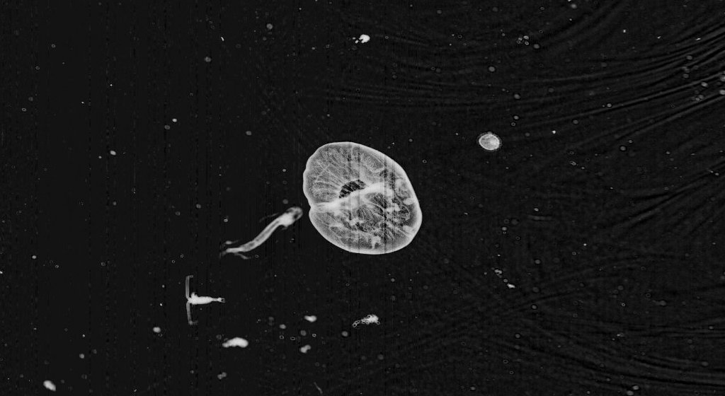
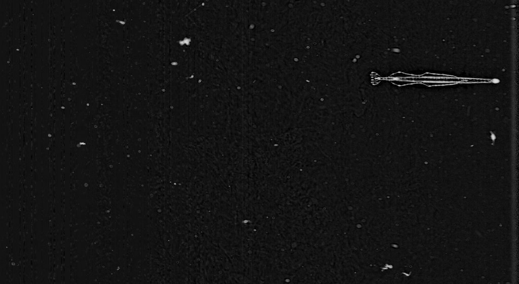
A year ago, we announced Plankton Portal 2.0, which featured a more streamlined design, a simpler tagging interface, and most importantly, a whole new dataset. Since then, this new data from the Mediterranean Sea has spurred a lot of interest and plenty of new questions. Participants on the site were surprised by the difference in size of everyone’s favourite jellies, the Solmarisidae (Solmaris rhodoloma in California, Solmissus albescens in the Med), which are much larger! Siphonophores also seem more abundant there. And the Mediterraean data came with brand new categories of organisms to mark: nice and cute medusa ephyrae (i.e. baby jellies), elegant Pteropods and the elusive fish larvae.
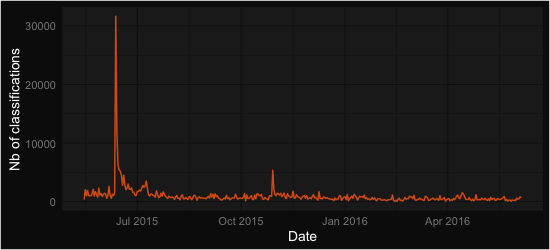
In total, as of last Sunday, 368,361 organisms were marked, on 50,519 distinct images. Through time, the classifications were marked by two peaks in activity: a huge one when the new version was announced through a mailing to the Zooniverse community (thanks everyone!) and another one when we pushed for 1,000,000 classifications in total, to celebrate Jessica’s PhD defense. When we zoom in, we see activity fluctuating around 1000 and now 500 classifications per day. This is still great (but coming back to 1000 would be even better! 😉 ).
time_series
time_series_zoomed


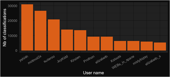
The top 11 contributors, all authors of over 5000 classifications each, are displayed below. If you made this top 11, we owe you special thanks (and probably a beer too). We hope you will stay interested and involved in this project. If you did not, you should really not be disappointed because all other volunteers still collectively account for 60% of the classifications; so you matter very much! Hopefully all of you will be happy to see some of the outcome of your work below.
who


Time for a bit of science! The most common classification was… nothing, empty, zero, zlich, zip… Well, you get the idea. Indeed, when we film the sea, we most often see nothing (nothing living at least). Even though we pre-selected potentially interesting frames for Plankton Portal (the ones having some kind of large object in them), about a third of your classifications did not contain any organism we were interested in. In real life, the proportion of dead detritus vs. living organisms is more around 95% vs. 5%, so our pre-filtering still avoided you a lot of blank frames! In terms of organisms, the 10 most abundant are shown in the figure below.

what


Doliolids, Copepods, and Radiolarian colonies dominate the rest. We immediately noticed, when we shot the images, that Doliolids were particularly abundant. Those organisms are very effective filtering machines and they may therefore have an impact on the density of smaller organisms, in particular unicellular algae. The relative abundance of Copepods and Radiolarian colonies is to be interpreted carefully: Radiolarian colonies can be large and span several frames (therefore increasing the total count) and Copepods are likely under-estimated because we mostly see the larger ones with ISIIS, and they are not the dominant ones in the Mediterranean. Still, it echoes nicely a recent Nature paper by Tristan Biard (a contributor to PlanktonPortal’s talk, under the username Collodaria), which showed that Rhizaria (a large taxonomic group to which Radiolarians belong) can be equivalent in biomass to Copepods, who were previously thought to largely dominate the plankton. These findings were also based on in situ images, because these fragile Rhizaria cannot be collected with nets.


Finally, the images in the Mediterranean were collected along transects (i.e. straight lines) perpendicular to the shore. We were interested in how organisms were distributed along a gradient between coastal and open ocean conditions. In the plots below, the coast is on the left, the open ocean on the right and the vertical direction is depth (top: surface; bottom: 100 m depth). So you basically see a “slice” of water along which ISIIS undulated. The size of the dots is proportional to the number of classifications recorded. You can immediately notice that Doliolids (first plot) are concentrated near the surface, and fish larvae (second plot) even more so! This is a surprising finding for fish larvae, which sometimes ended up in concentrations of over 10  individuals per cubic meter, a number much higher than what was previously observed elsewhere, with conventional plankton nets.
distrib_doliolid_w_icondistrib_fish_w_icon
Radiolarian colonies, on the opposite, tend to be concentrated in mid water (see figure below). Within this messy picture, some structure seems to emerge. Indeed, the white lines on top of the plot are contours of the concentration of Chlorophyll A in the water (i.e. of the amount of unicellular algae). If you look carefully, you will see that those lines are moving up, towards the surface, as we travel offshore (from left to right on the plot). This is actually well known in this region. What is interesting is that the radiolarians seem so follow the same pattern, and that higher concentrations of colonies sit on top of this high Chlorophyll region. Something is definitely going on between these two!
distrib_radiolarian_colonies_w_icon
That’s it for now — thanks again to everyone for this wonderful year of activity! We apologise for not being as active as we would like to be on Talk. To that end, we thank the active moderators who take over this important responsibility. And finally, we thank Zooniverse for the great opportunity and community they created. Now, on to next year!

Fantastic Find Friday Take 3!

Hey plankton hunters!  Welcome to our 3rd round of Fantastic Find Friday here at Plankton Portal.  There have been so many awesome finds on the site and we picked 5 this week for you to check out.  If you see something really neat on the portal than tag it with #FFF so we can check it out for use on the blog.  Here we go!

Physonect Siphonophore— #Sipho #Corncob

http://talk.planktonportal.org/#/subjects/APK0000iu4

51d1bd8e3ae74008a4006d0e

This is a stunning capture of a physonect siphonophore who seems to be waving hello to ISIIS as she passes by.  Like all siphonophores, this guy here is a colonial organism comprised of many individual animals or ‘zooids.’  Each zooid is specialized and distinct, but work together so closely that they more resemble a single organism than a colony of animals.  On display here are the branching tentacles used for foraging and the swimming bells that resemble a corncob.  This one is a stunner!

Lobate Ctenophore — #Lobate

http://talk.planktonportal.org/#/subjects/APK0000l30

51d1bd903ae74008a4007882

This is a really neat capture of a lobate ctenophore (Ocyropsis maculata), showing off the feature that gives this guy his name.  In this image you can see clearly the internal structure and the striated texture of his muscular, gelatinous body.  Lobate ctenophores swim lobes forwards by beating the ciliated comb rows situated on the opposite (aboral) end.  The one depicted here would be swimming towards us and to the left.  I wonder if larvacean is on the menu?

Chaetognath — #Arrowworm

http://talk.planktonportal.org/#/subjects/APK0000hpr

51d1bd8d3ae74008a4006758

Looks like an arrow shot by some undersea archer, right?  Arrow worms, or chaetognaths, are carnivorous marine worms belonging to the Phylum Chaetognatha.  They are notoriously ferocious predators that hunt other plankton with the help of hooked ‘grasping spines’ that flank the mouth.  Chaetognaths have fins for propulsion and steering—you can see all of them really well in this capture!  While these fins superficially resemble those of a fish, they are not related evolutionary and are structurally very different.

Calycophoran Siphonophore — #Rocketship #Triangle

http://talk.planktonportal.org/#/subjects/APK0000k4m

51d1bd903ae74008a40073a5

I bet NASA would get a lot more funding if they built space shuttles that looked like this!  This beautiful capture of a siphonophore really looks to me like some sci-fi monster a (horrified) astronomer might see in a telescope!  Don’t worry though, this guy is just a couple of cm’s long and probably couldn’t hurt you if he tried.  Just like the physonect siphonophore above, this guy is a colonial organism and would therefore be more appropriately referred to as guys.  The tail, or stem, on display here contains two developmental stages of siphonophore simultaneously—both the medusa and polyp stages.  Unlike most cnidarians that alternate between these stages generationally, this guy chooses to have them coexist within the same colony.  If you look closely you can see them bickering over who is the prettiest!

Calanoid Copepod — #Copepod

http://talk.planktonportal.org/#/subjects/APK00005l6

51d1bd843ae74008a4001c98

This copepod is making a heart with his antennae! Do you think he might be in love?  There is some 13,000 species of copepod in the world and they are a crucial component of plankton communities and global ecology in general.  It has been suggested that copepods may comprise the largest animal biomass on the planet! Many species of marine life, large and small, rely on these guys as their main food source, including whales and seabirds.  Looks like this guy here is a lover not a fighter!

Looking forward to next time !

Why do we need Citizen Science?

In many fields of science, new technology is leading to unprecedented data production. This, in turn, requires extensive analysis with minimal sub-sampling to detect as much detail as possible. In biological oceanography, imaging systems have become more useful with increasing computer speed and storage capabilities, and image data address some of the fundamental problems with traditional sampling methods that are destructive to fragile organisms (i.e., jellyfish and marine snow). On a given tow with our system, the In Situ Ichthyoplankton Imaging System (ISIIS), we produce approximately 400,000 images in 7 hours with many different species across a range of sizes present in each image (500 μm to 13 cm). This is an incredible amount of information that would take years for one person to fully analyze. When we are out at sea, we typically sample for WEEKS and come back to land with millions of images. Computer algorithms can perform basic tasks of extracting specimens that look similar, but human brains are extremely adept at interpreting an organism in 3D and providing context in the image data that a computer cannot. The amazing abilities of people to recognize patterns that computer algorithms may see as unimportant cannot be underestimated.

Shrimp photograph taken from under a microscope

Shrimp photograph taken from under a microscope (photo credit: Cedric Guigand)

Another reason we are using Citizen Science is so that you, the citizen scientist, can participate in the process of discovery. After all, most oceanographic research is funded at least in part by taxpayer money, and these novel plankton images combined with Citizen Science are a great way to engage those who fund the research. We think it is far more effective to cultivate interest in science through the discovery process itself, rather than the production of jargon-filled reports and papers only understood by other oceanographers (don’t worry, those will come later). In addition, this online format provides an opportunity for us to educate people about life in the oceans, potentially inspiring the next generation of ocean scientists. With Citizen Science, there is the potential for new discoveries arising from simply allowing many people to look at the images.

This larva of a deep water shrimp was captured in the Gulf Stream near South Florida (Photo credit: Cedric Guigand)

This larva of a deep water shrimp was captured in the Gulf Stream near South Florida (photo credit: Cedric Guigand)

We believe our research with ISIIS is particularly applicable to Citizen Science and the process of discovery because this new imaging technology provides a huge amount of data and a unique glimpse into ocean life. I have spent the last 5 years of my graduate school career at the University of Miami examining hundreds of thousands of plankton images, and every time I flip through the images, I always have the feeling that I could see something that no human has ever seen before. I try to instill this sense of wonder and hope for discovery in all people that work with the images, because when you see something interesting, like an elaborate siphonophore or a dense patch of copepods, you are likely the first person to see that species in its natural environment. When we get enough eyes on these images and discussions facilitated through the Plankton Portal website, the sky’s the limit for the discoveries that can be made with Citizen Science!

How do we get the images?

nice isiis3

Since the 1800’s, plankton has been studied and collected using simple nets with very fine mesh. The process of analysis of these plankton ‘samples’ is tedious, labor intensive, confined to laboratory, and can only be done on relatively small areas of the ocean. By sampling in this manner, it is difficult to get a good understanding of how planktonic organisms are distributed and how they interact with each other. Yet plankton represents a very important part of a global system feeding larger animals like fish, whales and many others. The In Situ Ichthyoplankton Imaging System (ISIIS) is one of a few systems in the world capable of improving the way we study plankton to better understand their life and function in the marine environment. Instead of using an actual net to capture plankton, ISIIS captures the images of the organisms and information about their immediate surroundings. ISIIS samples continuously, resulting in a collection of digital images that record the exact location of the various plankton organisms in relation to each other and the environment in which they live. Further, the images are recorded onto a simple hard drive instead of slurry of plankton all mixed together in a sample jar with formaldehyde (yech!).

IMG_0873

ISIIS is an underwater imaging system developed to capture real time images of plankton that are relatively rare, small, and fragile such as fish larvae and delicate gelatinous organisms (like jelly fish). ISIIS is composed not only of a macro-camera system with its own illumination but it also is integrated into an underwater vehicle with a variety of additional sensors to measure the depth, salinity and temperature of the water, as well as such properties as dissolved oxygen, light level, and even how much chlorophyll a (measure of primary production) is present. Together, the camera and sensors provide detailed profiles and tracks of what plankton are where and what the ocean environment around them is like.

isiis schematic

The vehicle, and associated imaging system and sensors, moves up and down through the water column using side-mounted, user-controlled dive fins (like an underwater glider) while being towed behind an oceanographic ship moving at 5 knots. The vehicle frame is divided into four compartmentalized enclosures with imaging and optical equipment seamlessly integrated into ISIIS’s ventral housings and environmental sensors and electronics in the dorsal housings. ISIIS is designed to undulate in a zigzag fashion between the surface and a maximum depth of 200 meters.

isiis2

The ISIIS system utilizes imaging technology very similar to an office scanner flipped on its side. The imaged parcel of water passes between the forward portions of two streamlined pods where it is “scanned” and transformed into a continuous image. The resulting very high-resolution image is of plankton in their natural position and orientation. When a sufficient volume of water is imaged this way, quantification of concentration (individuals per unit volume) and fine scale distribution is possible. ISIIS is capable of imaging a maximum of 162 Liters (43 gallons) of water per second (when moving at 5 knots) with a pixel resolution of 70 µm (the thickness of a human hair).

The imaging data and associated oceanographic data are sent to the surface ship via a fiber optic cable and recorder onto a main computer for later viewing and analysis.