http://www.dosits.org/people/investigateanimals/measureplankton/

Artwork by Jean-Olivier Irisson
Great news! We are working on translating Plankton Portal in French with our
French Collaborators: Fabrice Not from the Station Biologique de Roscoff and Jean-Olivier Irisson from the Observatoire Océanologique and Station Zoologique de Villefranche-sur-mer. “The idea is also to get some interest from French schools to develop a curriculum around Plankton Portal” Dr. Irisson explains. Stay Tuned.
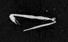
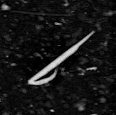
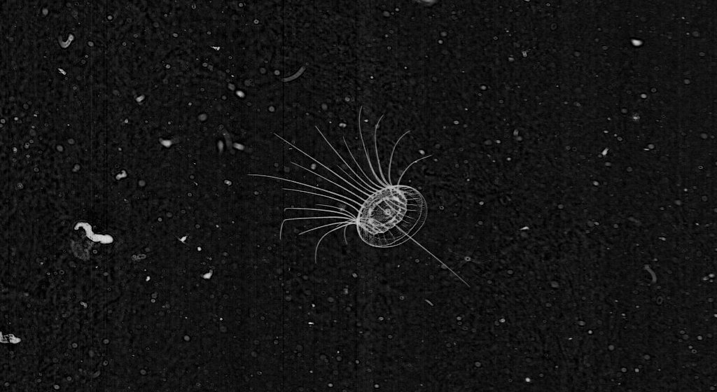
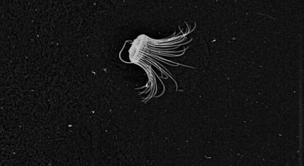
Of all plankton groups, probably most is known about the copepods. They represent a critical link in the food chain and are consumed by diverse animal community ranging in size from small fish, chaetognaths, and ctenophores all the way up to large whales (the right whale is a voracious copepod feeder). Because of their small size and importance as food, copepods are affectionately known as “the rice of the sea.” Copepods are effectively captured by plankton nets because they have hard exoskeletons, and scientists have good estimates of their abundances and distributions. Although copepods are all relatively small (0.5 mm – 5 mm in length), they comprise over 200 families and 10,000 different species.

Examples of typical copepods. Note to the two large appendages on the top of the head with small sensory hairs
Copepods consume both phytoplankton and microzooplankton in two different ways: suspension feeding and raptorial feeding. Suspension feeding is relatively passive and performed by beating small appendages that draw a current through a feeding chamber. Copepods then select which particles encountered are food and discard others. Raptorial feeding is used to actively capture prey. Many copepods have small sensors on their first appendages to detect water disturbances produced by prey and also predators. They can use these relatively large appendages to “hop” through the water and capture an unsuspecting prey item or to quickly escape a predator.
Copepod reproductive strategies vary greatly and are adapted towards the ability to withstand the variable conditions that characterize the ocean environment. For example, many copepod eggs have the ability to enter a phase of diapause where they remain viable on the bottom for several months or even years, only hatching with conditions are favorable (high concentrations of food). Some copepods carry their eggs, allowing them to develop a bit before releasing them into the water column. The timing of copepod reproduction is especially important for the life cycle of fishes because most fish larvae depend on the recently hatched copepod nauplii for food. If there are not enough copepod nauplii present when fish larvae are abundant, there could be mass starvation events causing few fish larvae to reach their juvenile stage. Because of this, the copepod life cycle is extremely important to fish populations and overall ocean ecosystem health.
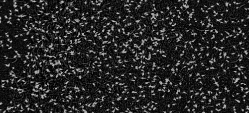
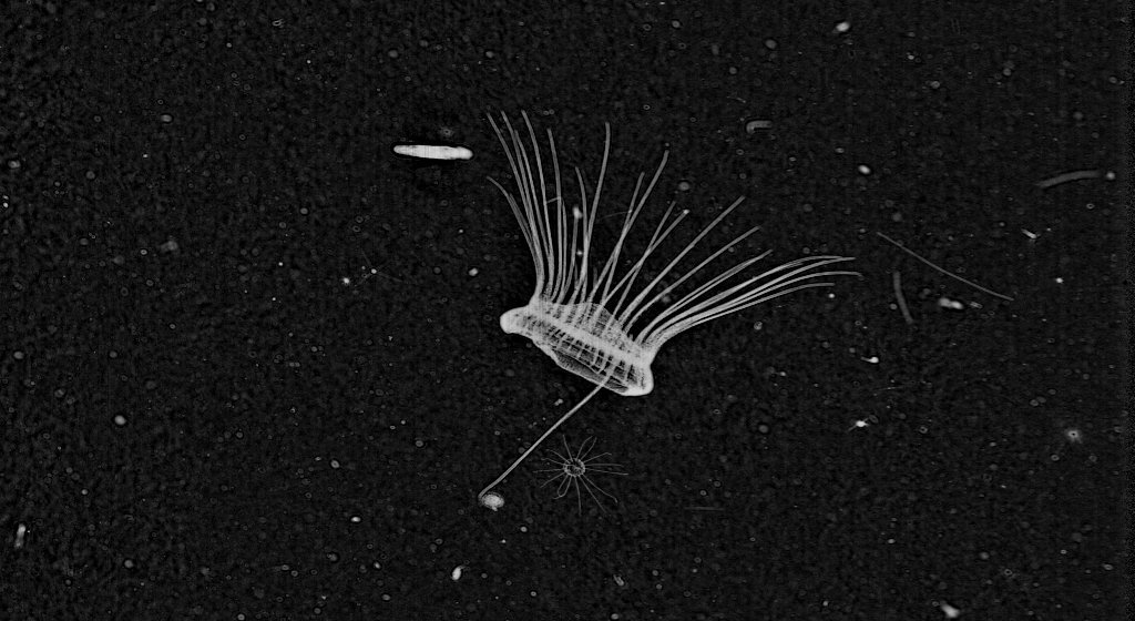
This image was taken from a thin layer near Stellwagen Bank offshore of Massachusetts, USA. Each one of the white particles is a copepod. The concentration of organisms in this image corresponds to ~400,000 individuals per cubic meter! That is some good eating for a right whale!
One of the most remarkable characteristics of copepods is their tendency to aggregate in discrete thin layers within the water column. Sometimes >90% of the copepod biomass will be confined these thin layers, which are a maximum of 5 m thick. ISIIS and other systems that sample on small scales are ideal for detecting these layers of copepods, and the function of the formation and dissipation of copepod thin layers is not well understood. Copepods have been shown to be attracted to strong changes in current direction and speed, potentially allowing them to feed at a faster rate within these zones (Woodson et al. 2005). The changes in environmental variables associated with aggregations of copepods are of great interest to marine ecologists. With your help, we can better understand how these extremely important organisms are distributed throughout our oceans!
References:
Johnson WS, Allen DM (2005) Zooplankton of the Atlantic and Gulf coasts: A guide to their identification and ecology. Johns Hopkins University Press. Baltimore, MD.
Woodson CB, Webster DR, Weissburg MJ, Yen J (2005) Response of copepods to physical gradients associated with structure in the ocean. Limnol Oceanogr 50:1552-1564
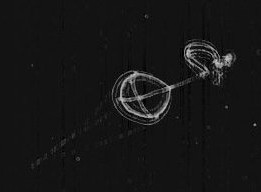
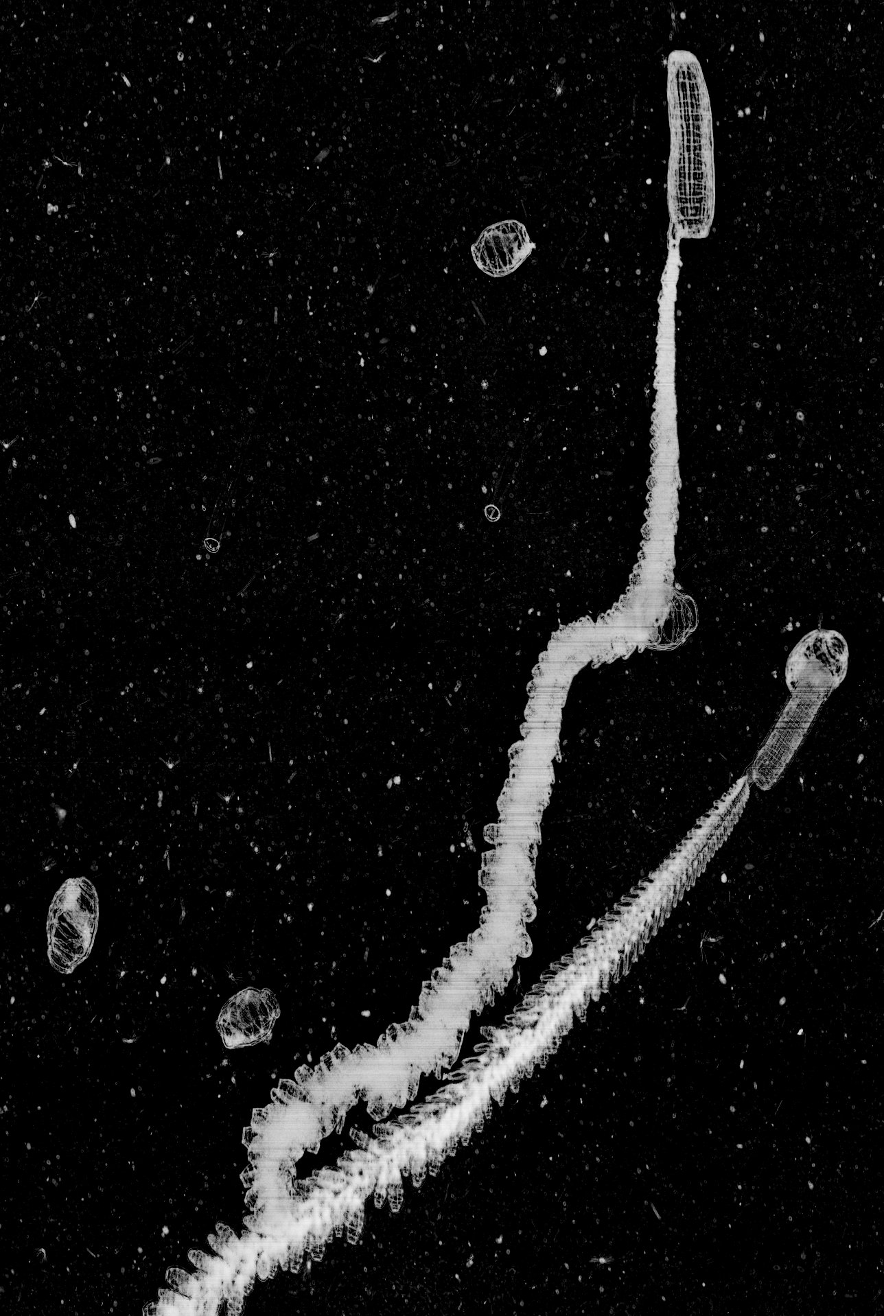
Pteropods are a group of organisms that we’re not focusing on because they are not very abundant in the Plankton Portal dataset. Nevertheless, you may have run across a few of those fascinating little creatures.
Pteropod, which means ‘wing-foot’ in Greek, is a group of free-swimming pelagic gastropods (snails). Officially, the word ‘pteropod’ is no longer used in taxonomy; it is a collective term which refers to two clades of gastropods—thecosome (shelled body) and gymnosome (naked body). Pteropods are quite unique because in order to adapt to life in the water column, their foot is modified into two wing-like flippers used for swimming. Their body size ranges from a few millimeters to several centimeters – so they’re easily imaged by ISIIS. They can be quite abundant in certain regions of the world’s oceans, and are typically found near surface waters.
The first group of pteropods, thecosomes, are also known as the sea butterflies. They have a pair of large ‘wings’ and swims by continually flapping them. Their body is encased in a delicate and translucent shell.The shell can be coiled, needle-like, triangular, and globed.
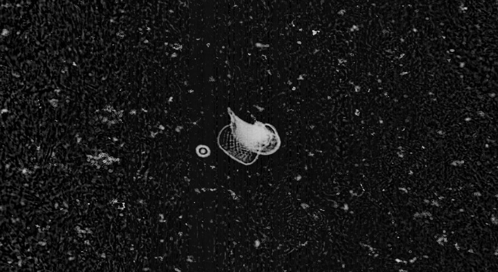
Thecosomes are omnivores. Their diet consists of diatoms, dinoflagellates, and zooplanktons such as copepods, tintinnids, and other gastropod larvae. They capture food by secreting a spherical mucus web several times larger than their body. Scientists believe that the use of the large size mucus web is to capture large, fast swimming prey, such as copepods. The web acts as a filter: particles that are too large for ingestion are removed. During feeding, the mucus web is suspended above the animal while the animal remains motionless below. Ciliary action draws back the web to the mouth and the whole web is ingested.

Thecosome reproductive biology is quite unusual. The animal first matures and functions as male. The male pteropod mates with another male and the sperm is stored until the animal changes into a female. When the animal turns into female; its male reproductive organs degenerate. The female lays fertilized floating egg mass that later hatch into swimming larvae (veliger).
When a thecosome dies, its shell sinks to the bottom of the sea and forms sediment called pteropod ooze. The shell is composed of aragonite, an unstable form of carbonate mineral. Anthropogenic ocean acidification is one of the challenges that pteropods face. The increase of anthropogenic carbon dioxide level in the atmosphere reduces pH and carbonate ion concentration in the ocean, thus decreasing the calcium carbonate saturation level. As a result, the production of biogenic carbonate becomes more difficult. Overall, they have a hard time secreting their protective shell because of ocean acidification.
The second group of pteropods, or gymnosomes, are more commonly known as sea angels. They have much smaller wings which appear as side lobes. They are more robust and lack a shell. Unlike their thecosome relatives, gymnosomes are carnivores. They are active hunters and exclusively prey on thecosome pteropods. A combination of hooks and a toothed radula are employed to extract the flesh from the thecosomes’ shells.
The reproductive anatomy of gymnosome pteropods is similar to thecosomes pteropods. The only difference: the male reproductive organs do not degenerate in females. Gymnosomes has two distinct larvae forms. Eggs are hatched into shelled veliger. The veliger metamorphoses into a shell-less polytrochous larvae. The polytrochous larvae are initially wingless and movement is achieve by three ciliary bands. They gradually grow wings and lose the ciliary bands as they become adults.
Here is a very nice video about Pteropods.
Plankton Chronicles Project by Christian Sardet, CNRS / Noe Sardet and Sharif Mirshak, Parafilms. See Plankton Chronicles interactive site: planktonchronicles.org
Hello everyone. We have a special “behavior” Fantastic Finds Friday (FFF) today! These frames were selected from your posts to illustrate the power of the human eye to detect rare and unusual phenomena. The frames selected here may not be the most beautiful you have seen so far, but the story behind them is fascinating and could not be told without the help of our citizen scientists.
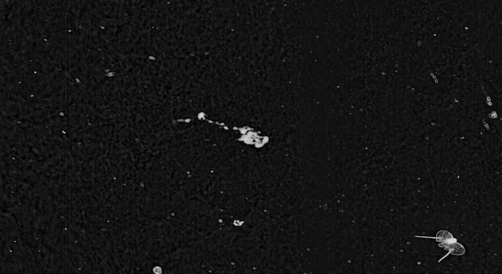
Here is great shot of a larvacean (also known as an appendicularian) getting spooked by the movement of ISIIS. Larvaceans are known to escape from their mucous house if threatened by a predator. Unfortunately the house can’t be used again, and they will start secreting a new house once the threat is passed.

Arrow worms (chaetognath) are voracious predators capable of engulfing prey as big as their own body. In these images, you can see an arrow worm catching a larvacean and the other grasping what appears to be a copepod. Their mouths resemble a crown of spikes ready to impale any unlucky prey. Chaetognaths also prey on fish larvae.
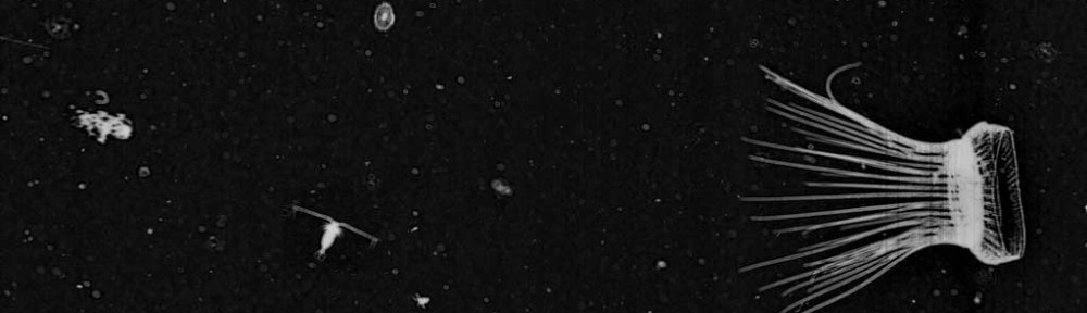
These two medusae just snagged a larvacean house. Accident or deliberate attempt to feed on these poor guys? The long trailing tentacles act like a sticky fishing net that retracts to bring in the catch of the day.
These Solmaris seem to be reaching for something (one tentacle pointed opposite to the others). Solmaris have been seen feeding on other jellies – even large siphonophores! They swim with their tentacles forward to maximize the chances of catching a prey. they then move the item to their mouth with one tentacle (like an arm almost).
No, these are really two different frames! Amazing consistency in posture isn’t it? And look at these two tentacles reaching out – sensing their environment? Hoping to encounter a tasty prey item? If we detect enough of these organisms, we could try to investigate at which time or location they behave this way. This could be a really interesting project!


So if you see something interesting like these example or suspect some interaction is at play in one of the frame use the hashtag #behavior. Remember to mark frames you want considered for future Fantastic Finds Friday posts with #FFF. Thanks, and keep up the good work!
Greetings plankton enthusiasts, new and old! My name is Jenna Binstein and I recently graduated from undergrad at the University of Miami. I enjoyed my time there so much though, that I signed on for another year as a graduate student! Part of what made my undergraduate years so fulfilling and worthwhile was my work in the lab with Dr. Cowen, Jessica, and the rest of the ISIIS/plankton team. Before I go into more detail about my work there, let’s take a quick look at how I found my way into the marine sciences.

It all started when I got my SCUBA certification as a freshman in high school. After my first open water dive I was hooked. I knew I had to learn all there was to know about marine science. At first, I thought I wanted to study the “big” stuff: dolphins, sharks, or turtles. I had seen jellyfish on SCUBA dives before, but I always considered them pests. I never thought as I applied and enrolled at UM that I would find such passion in studying some of the smallest organisms in the ocean, and learn just how important and collectively “big” they actually are.
Basically, my journey with plankton started when I met Jessica and Adam and began helping them with their respective dissertation research. I started learning to identify zooplankton, just as you all are learning to do via Plankton Portal! I started getting comfortable with the images from ISIIS, and eventually began to develop my own interest and senior thesis project with mentorship from Jessica. I decided to begin looking more closely at Appendicularians. Very little is known about these guys and their unusual mucous housing. So I spent a long time quantifying Appendiularians by size, classification, and whether or not they were inside a mucous housing when I saw them. The goal was to be able to identify an existing relationship between depth and whether or not an Appendicularian was found in its housing. I briefly looked at other factors as well, such as frontal dynamics, size, and classification and then saw if these related to an Appendicularian being in or out of its house. Although I completed my senior thesis, the work is not over; as there is still so much more I can pull from the data! Yet overall, I learned so much about Appendicularians and their role in the oceans, and I will definitely share as much of that as I can with all of you on some later blog posts relating specifically to the Appendicularian. In the meantime, I hope to continue learning all I can about Appendicularians and other gelatinous zooplankton during my time with the help of ISIIS, Plankton Portal, and UM.
Until my next post, happy jellyfishing everyone ≡≡D

Jenna Binstein, B.S.M.A.S., is a student in the Masters of Professional Science program at the Rosenstiel School of Marine and Atmospheric Sciences (RMSAS), University of Miami. You can reach her at jbinstein [at] rsmas.miami.edu.
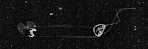
Salps and doliolids (class Thaliacea) are interesting animals because they are in the phylum Chordata, which includes all animals with a notochord during development (e.g., humans, fish, cats), but thaliaceans have a vastly different appearance and feeding strategy compared to most vertebrates. A salp or doliolid body is essentially a giant pumping muscle that forces water through a mucous net filter that collects phytoplankton and is ingested periodically. Both groups have limited mobility, with salps using muscular contractions to scoot through the water, while doliolids use tiny beating cilia to propel themselves.

A salp in the process of forming a new chain of clones for asexual reproduction (see white coil)
The life history of salps and doliolids is remarkable and complex. Similar to plants, their life cycle alternates between sexual and asexual generations. The solitary phase reproduces asexually by budding off clones of itself. On salps, a chain of these clones develops on the solitary animal that is then released and reproduces sexually with other salp chains. The chains first mature as female and then change sexes to become male when they are larger! These chains release small solitary salps that then begin asexual budding once they are a certain size. Doliolids on the other hand produce short-lived tadpole larvae that are not seen in salps. When you consider that a chain of salps contains an average of ~28 individuals, it is no surprise that these organisms are capable of extremely fast reproductive rates and can double their populations in hours (Heron 1972). Some scientists think their remarkable reproductive rates can overwhelm other phytoplankton grazers, which could explain the fact that large salp aggregations are often associated with low biomass of other grazers (Alldredge and Madin 1982).
Doliolids imaged offshore of Monterey Bay showing asexual budding
Because of their ability to reproduce quickly, salps are often very abundant near steady supplies of phytoplankton, such as at ocean fronts (zones where two water masses with differing physical properties meet) and eddies (Deibel and Paffenhöfer 2009). However, these organisms cannot tolerate extremely dense aggregations of phytoplankton because their mucous filters will become clogged with prey, which severely decreases their feeding efficiency. Salps and doliolids can “bloom” like other jellies, and when these blooms die off the dead salp bodies can export a large amount of carbon into deeper waters. Because of salps and doliolids close evolutionary relationship to vertebrates, scientists are also very interested in their developmental biology. Scientists are trying to use salps as a model organism to study the development of complex nervous systems in all vertebrate animals (Lacalli and Holland 1998).
Check out this video from Plankton Chronicles on these remarkable animals!
Plankton Chronicles Project by Christian Sardet, CNRS / Noe Sardet and Sharif Mirshak, Parafilms. See Plankton Chronicles interactive site: planktonchronicles.org
References:
Alldredge AL and Madin LP (1982) Pelagic tunicates: Unique herbivores in the marine plankton. Bioscience 32:655-663
Deibel D and Paffenhöfer GA (2009) Predictability of patches of neritic salps and doliolids (tunicata, thaliacea). J Plankton Res 31:1571-1579
Heron AC (1972) Population ecology of a colonizing species: The pelagic tunicate Thalia democratica – I. individual growth rate and generation time. Oecologia 10:269-293
Lacalli TC and Holland LZ (1998) The developing dorsal ganglion of the salp Thalia democratica, and the nature of the ancestral chordate brain. Philosophical Transactions of the Royal Society B: Biological Sciences 353:1943-1967
We are nearing the end of Friday, so apologies that this post is late! Hopefully it will be enjoyable for you weekend warriors! By the way, did you see that we are almost at 200,000 classifications?! I am so impressed by this amazing group of citizen scientists that make Zooniverse projects a success, particularly this one. THANK YOU.
We are going to use FFF to point out some amazing pictures that you guys have identified and called to our attention in the last week+, and also to clarify some confusion on a tricky category.

http://talk.planktonportal.org/#/subjects/APK00003ui
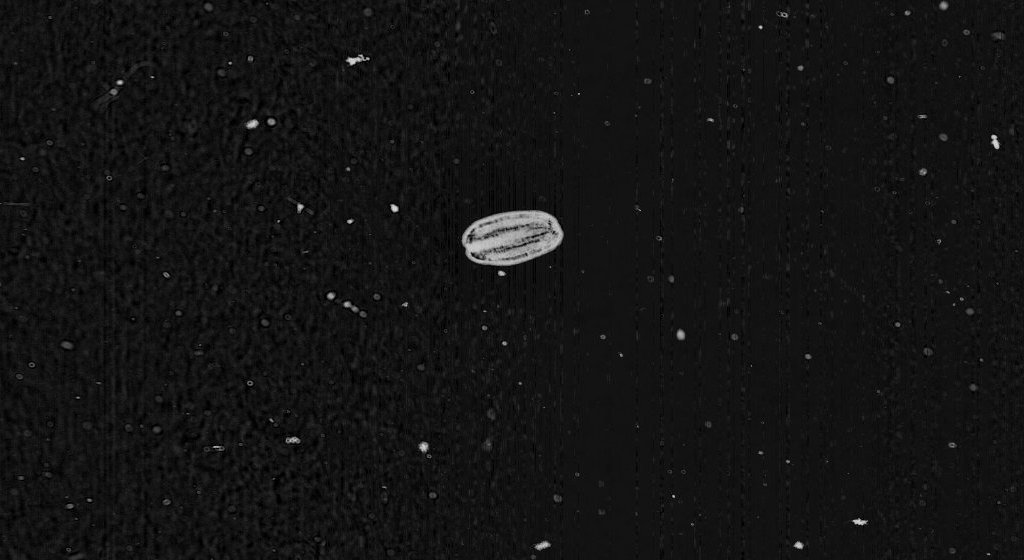
Cydippid ctenophore – #cydippid
This is a type of comb jelly, called a cydippid ctenophore. We think that this organism is Hormiphora californiensis or a relative. It has a egg shaped body with two tentacles, which are typically extended (for feeding), but also can be retracted into the sides of its body.
The relative of Hormophora californiensis is Pleurobrachia bachei, the sea gooseberry. Check out the following video of P. bachei feeding on some brine shrimp:
Here is another video of P. bachei from the Vancouver Aquarium:
For every easily classified cydippid ctenophore there is also other cydippids that are more difficult to classify by users. These ctenophores include Mertensia and Haeckelia beehleri, which are also cydippids but have their tentacles withdrawn. See below:
http://talk.planktonportal.org/#/subjects/APK0000yb7
This is also a cydippid ctenophore – but it has its tentacles withdrawn.
To add some more complication to the matter, there are also some lobate ctenophores, like the one below, whose young have a cydippid-like phase.

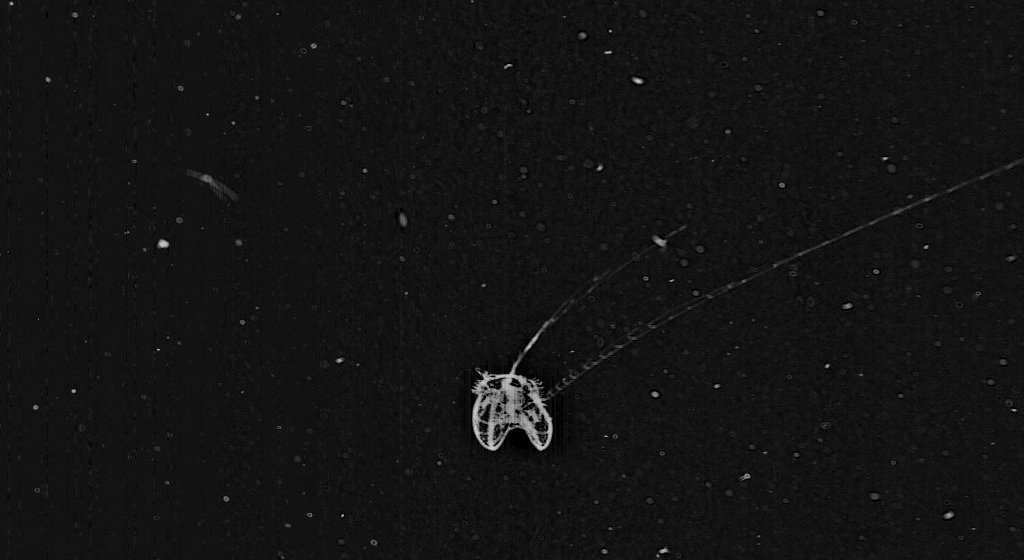
http://talk.planktonportal.org/#/subjects/APK0000kvy
Lobate ctenophore – #lobate
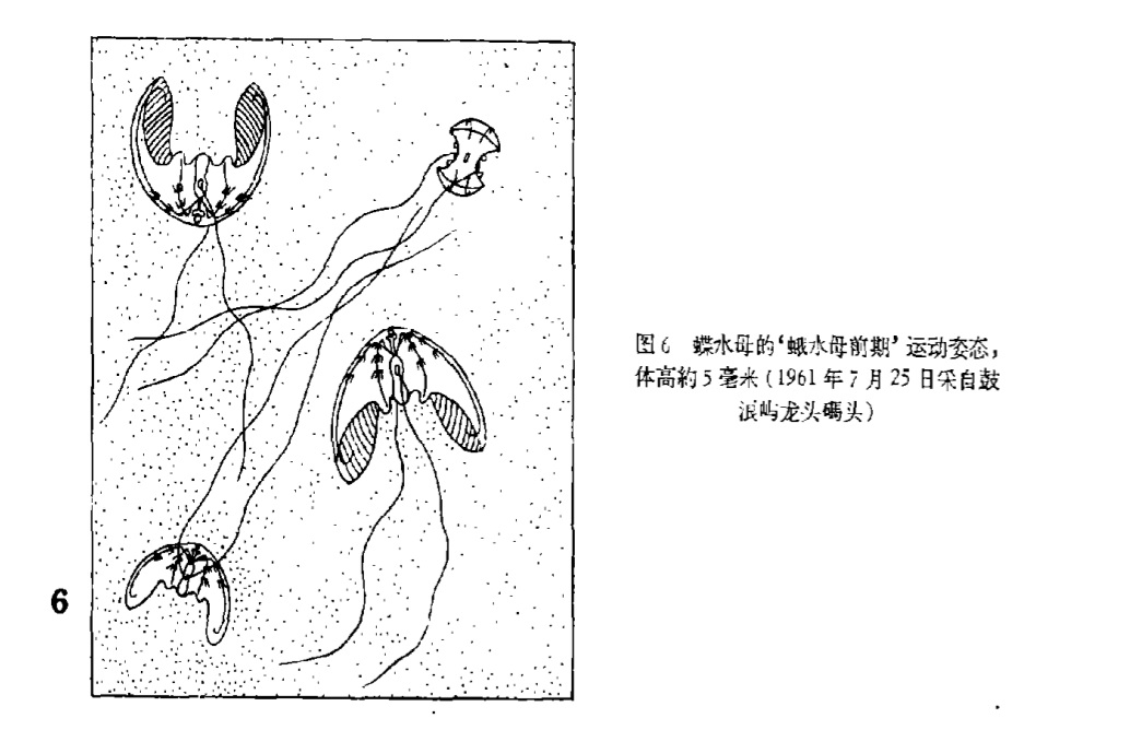
This is a beautiful shot of an adult lobate ctenophore, most likely the species Ocyropsis maculata. However, their young have this cydippid-like phrase. There has been one paper that published a drawing of the development of Ocyropsis. It was published in 1963. I had to email all around to get a copy, and when I receive it, I see that it’s in Chinese. Fortunately, they had great drawings that helped me.
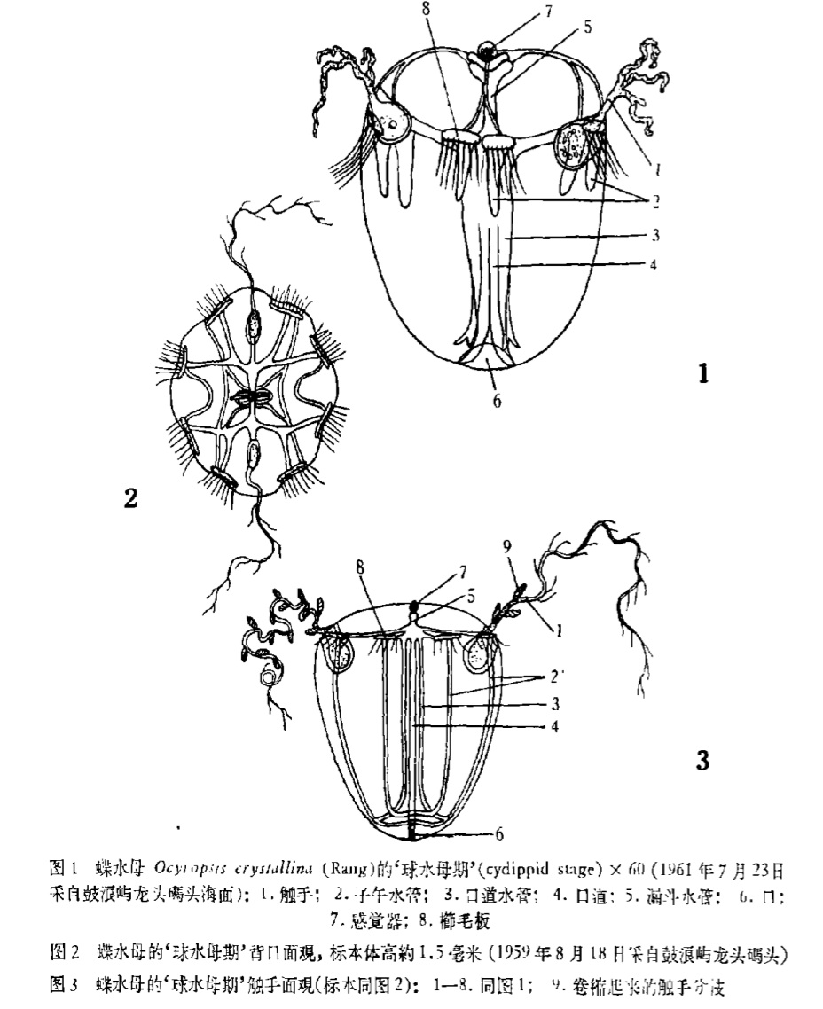
Chiu SY (1963) The metamorphosis of the ctenophore Ocyropsis crystallina from Amoy. Acta Zoologica Sinica 15:10-16
If anyone can translate the Chinese, let us know! But otherwise, just look at the cool pictures. There are a couple different stages of lobate ctenophore development, and the cydippid stage is one of the earliest stages. We definitely see this stage in our images. See below:
http://talk.planktonportal.org/#/subjects/APK0000y4e
Cydippid-phase of young Lobate Ctenophore
Officially, we want you to make this as a #lobate. BUT, we also know that these are incredibly confusing because these ctenophores have tentacles. So, we understand if you get these mixed up. In our data cleanup, we will end up checking the classifications of the small cydippids and lobates to make sure that they are classified correctly. Also, please know that if you do mix these classifications up, we will at least know that it’s a ctenophore! That’s more information than we had previously. So, anything is helpful.
THAT’S ALL FOLKS! Thanks for reading. Remember to tag images you want considered for Fantastic Finds Friday with the hashtag #FFF. And as always, thanks for classifying! We are currently at 191,968 classifications. So very close to 200,000!
Once again, we come on here thanking all of you immensely for your efforts in classification on this blog! We have nearly reached 200,000 classifications!!
We are at (drumroll please) 191,736 classifications!
Will you help us meet 200,000?

