100,000! Thank you to our top classifiers

Plankton Portal reached 100,000 classifications on Friday! In honor of our 100,000th classification, we’d like to publicly thank our top classifiers since our launch.

Solmaris rhodoloma, our Plankton Portal mascot

Solmaris rhodoloma, our Plankton Portal mascot

The top ten are (drumroll please):

1. yshish
2. lynb
3. elizabeth
4. Ingolme
5. KarenLK
6. localwormguy
7. cnorvalk
8. charcinders
9. VBear
10. Collodaria

The next ten classifiers are:
Lounalune, darylh69, mlmuniz, shocko61, SandersClan, jim24, Sheepdog, Steve3455, tadaemdg, and csams

A huge THANK YOU to everyone who has helped us reach 100,000 + classifications!

Love,
The Plankton Portal Science Team

Fantastic Finds Fridays: Week 2! #FFF

We are at the end of week 2 and we pulled out some of the best finds from this past week. As a reminder, every Friday we will post a selection of Fantastic Finds. If you think you have found something really great on Plankton Portal then tag #FFF and we will check it out for use on the blog. Thanks for tagging your favorites this week!
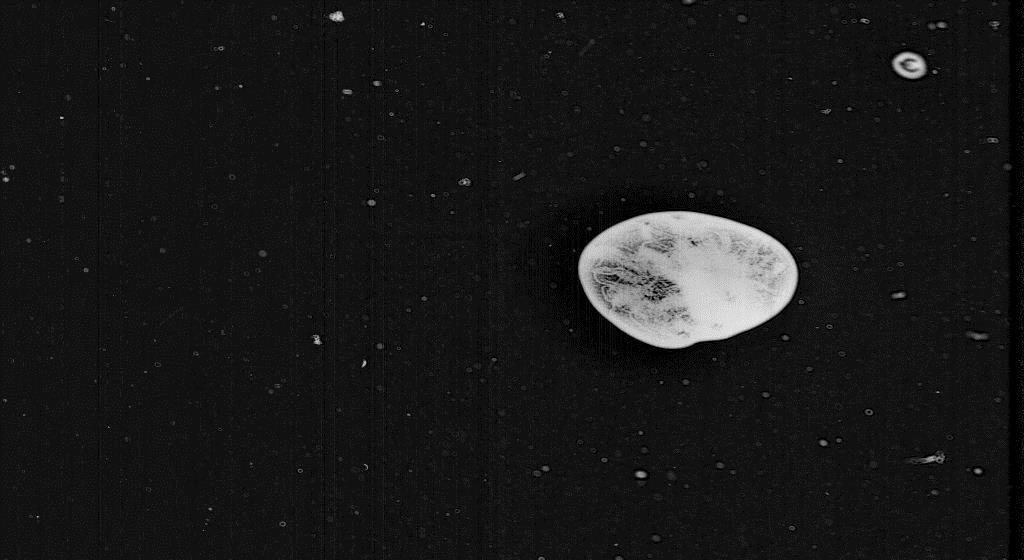
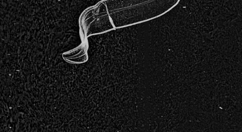
51d1bda03ae74008a4010740

Larval fish
http://talk.planktonportal.org/#/subjects/APK00015nq

Larval fish are actually considered part of the plankton, as fish in their early life stages will drift along in the oceanic environment. Because larval fish are relatively poor swimmers, they are under high predation pressure and more than 99% of baby fish that hatch from eggs will not make it! It’s a tough life. You might not know it from this site, but studying larval fish is a major component of our lab. Dr. Cowen has spent his career studying larval fish, their distributions, dispersal and population connectivity. In this particular study, we did not sample very many larval fish so we did not include it as one of the categories. However, we are incredibly interested whenever we see one so definitely tag the fish in the forum when you see any! #Larval #fish

51d1bd943ae74008a4009b1e

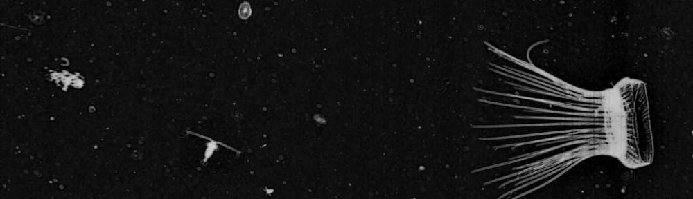
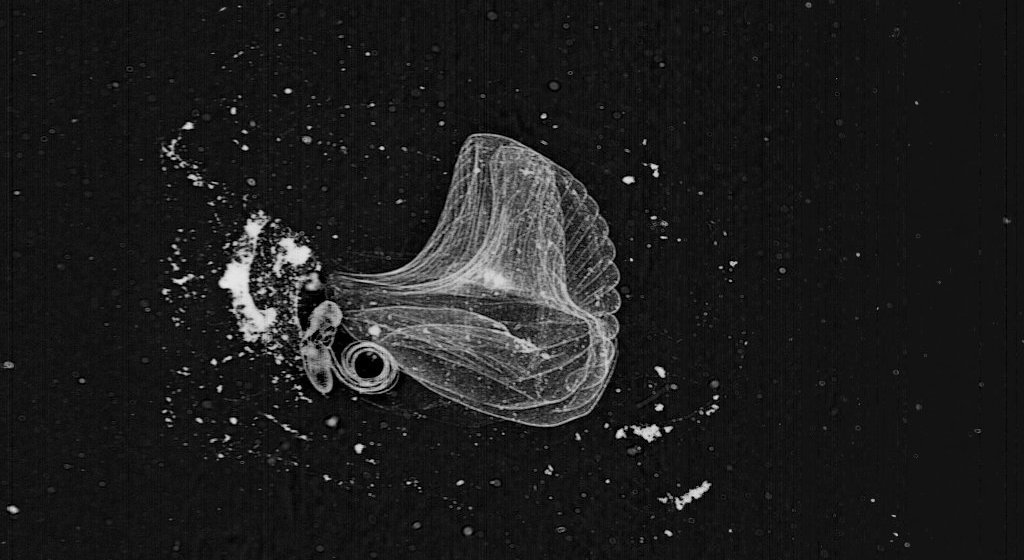
Liriope tetraphylla (#Medusae #4tentacles) with Arrow worm
http://talk.planktonportal.org/#/subjects/APK0000q5x

This is one of my favorite pictures from this week because what you see Liriope tetraphylla actually eating the arrow worm! Here one of his tentacles has brought up the arrow worm into the gastric peduncle (that’s the long thin appendage in the middle of the umbrella that looks like a handle). He appears to be holding the arrow worm in place while he eats his dinner. As far as I know, the only scientific study of what Liriope eats is from a paper by Larry Madin in 1988, published in the Bulletin of Marine Science, where he found that Liriope eats larvaceans, crustacean larvae, heteropods and juvenile fish. No one has reported that Liriope also eats arrow worms … until now.

51d1bd823ae74008a4000c10

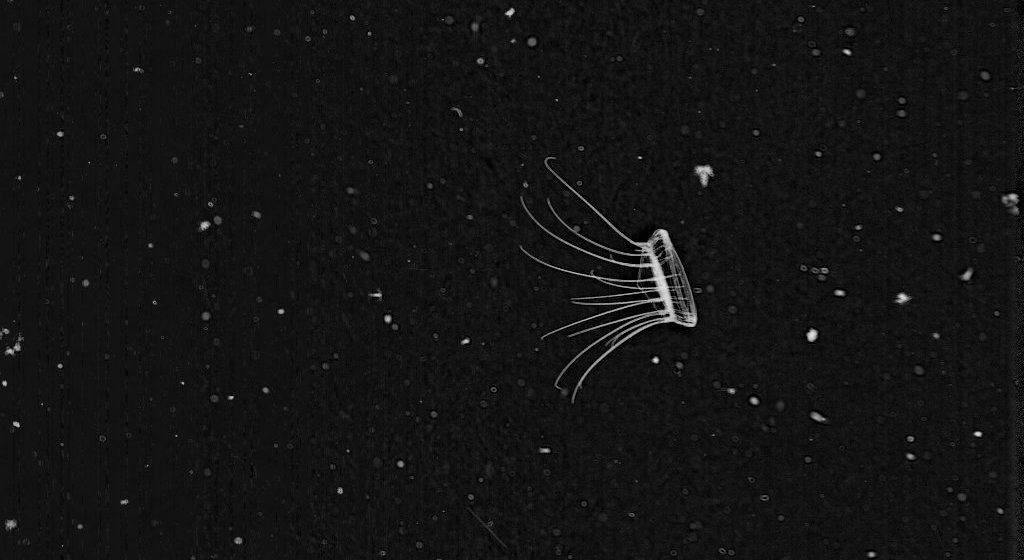
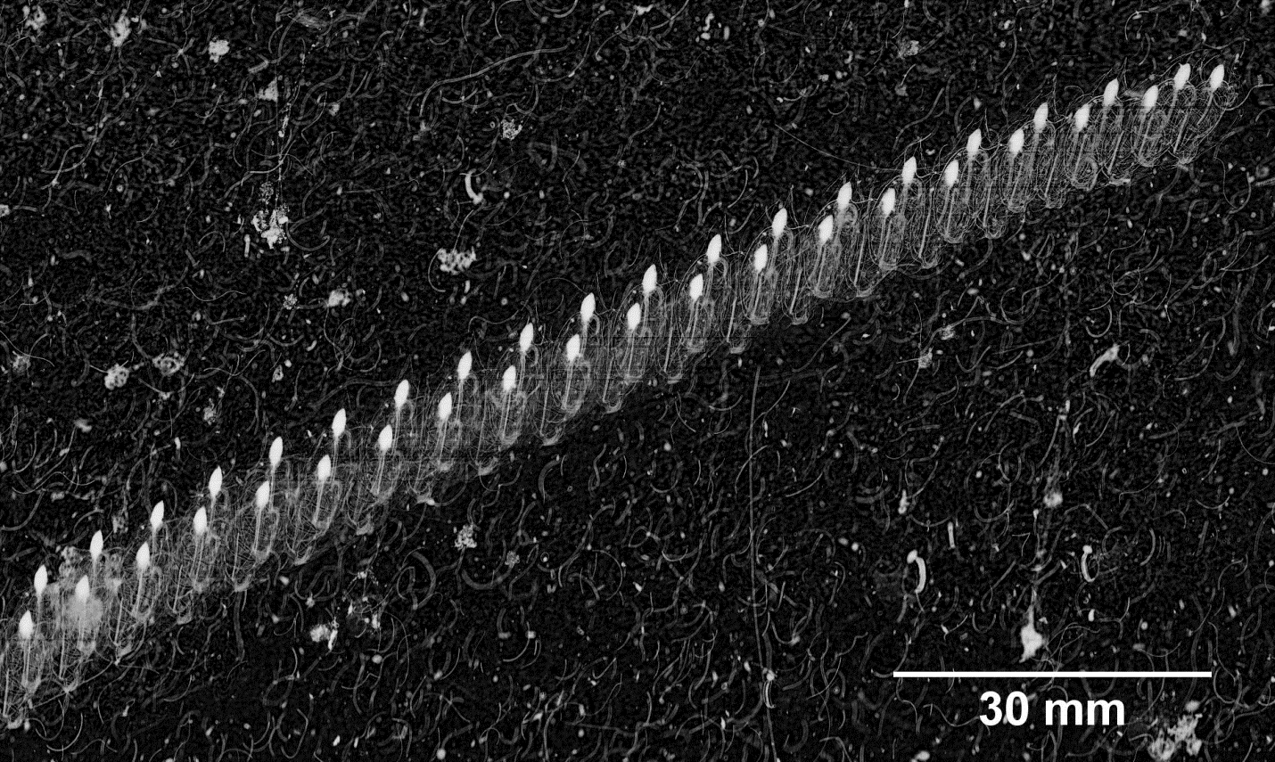
Sphaeronectes koellikeri – #rocketship #thimble
http://talk.planktonportal.org/#/subjects/APK00002cl

This beautiful creature falls within the broad group of jellyfish-relatives called the Siphonophores. Here you see this animal in a stunning feeding display. Though these guys are small and relatively inconspicuous, other siphonophores can get up to hundreds of feet long, and as a group are considered the deadliest predators in the ocean.  One fun fact: these rocketship siphonophores grow from the base of the stem towards the tail end. So the tail end of the stem is one of the oldest parts of the body. Sometimes you’ll even see small rocketships budding from the tail!

51d1bd833ae74008a4001258

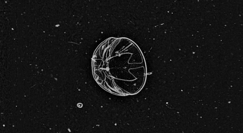
Radiolarian colony – #radiolarian #colony
http://talk.planktonportal.org/#/subjects/APK00003kq

We know that you’ve been frustrated by those small fuzzy round objects that invite classification but really aren’t supposed to be classified. Those are protists, a diverse group of eukaryotic microorganisms. One type of protist is the radiolarian, which are known for their glass-like exoskeleton, or “tests.” They are incredibly important in marine science because their tests are made of silica, which are preserved in marine sediments after they die and sink to the bottom of the ocean, and provide a record for paleo-oceanographic conditions, such as temperature, water circulation, and overall climate.

Radiolarians also form colonies. Colonial radiolarians are interesting because first, little is known about them, despite their abundance in the open ocean, and secondly, they are hosts to symbiotic algae that are modest but significant primary producers in the ocean. It has also been suggested that we are vastly underestimating the abundance of radiolarian colonies. Since primary production (photosynthesis, the conversion of sun energy into carbon) is the basis upon which all ocean life can exist, it’s incredibly important to understand who all the different primary producers are and how many of them are out there!


That’s all, folks. Thanks for reading, thanks for classifying, and remember: mark your favorites with #FFF for next week’s Fantastic Finds Friday!

Why do we need Citizen Science?

In many fields of science, new technology is leading to unprecedented data production. This, in turn, requires extensive analysis with minimal sub-sampling to detect as much detail as possible. In biological oceanography, imaging systems have become more useful with increasing computer speed and storage capabilities, and image data address some of the fundamental problems with traditional sampling methods that are destructive to fragile organisms (i.e., jellyfish and marine snow). On a given tow with our system, the In Situ Ichthyoplankton Imaging System (ISIIS), we produce approximately 400,000 images in 7 hours with many different species across a range of sizes present in each image (500 μm to 13 cm). This is an incredible amount of information that would take years for one person to fully analyze. When we are out at sea, we typically sample for WEEKS and come back to land with millions of images. Computer algorithms can perform basic tasks of extracting specimens that look similar, but human brains are extremely adept at interpreting an organism in 3D and providing context in the image data that a computer cannot. The amazing abilities of people to recognize patterns that computer algorithms may see as unimportant cannot be underestimated.

Shrimp photograph taken from under a microscope

Shrimp photograph taken from under a microscope (photo credit: Cedric Guigand)

Another reason we are using Citizen Science is so that you, the citizen scientist, can participate in the process of discovery. After all, most oceanographic research is funded at least in part by taxpayer money, and these novel plankton images combined with Citizen Science are a great way to engage those who fund the research. We think it is far more effective to cultivate interest in science through the discovery process itself, rather than the production of jargon-filled reports and papers only understood by other oceanographers (don’t worry, those will come later). In addition, this online format provides an opportunity for us to educate people about life in the oceans, potentially inspiring the next generation of ocean scientists. With Citizen Science, there is the potential for new discoveries arising from simply allowing many people to look at the images.

This larva of a deep water shrimp was captured in the Gulf Stream near South Florida (Photo credit: Cedric Guigand)

This larva of a deep water shrimp was captured in the Gulf Stream near South Florida (photo credit: Cedric Guigand)

We believe our research with ISIIS is particularly applicable to Citizen Science and the process of discovery because this new imaging technology provides a huge amount of data and a unique glimpse into ocean life. I have spent the last 5 years of my graduate school career at the University of Miami examining hundreds of thousands of plankton images, and every time I flip through the images, I always have the feeling that I could see something that no human has ever seen before. I try to instill this sense of wonder and hope for discovery in all people that work with the images, because when you see something interesting, like an elaborate siphonophore or a dense patch of copepods, you are likely the first person to see that species in its natural environment. When we get enough eyes on these images and discussions facilitated through the Plankton Portal website, the sky’s the limit for the discoveries that can be made with Citizen Science!

Amazing Plankton Videos

Hi all! we wanted to share this website, called Plankton Chronicles, with you. It is an amazing collection of plankton related mini videos. You’ll get to see some color videos of your favorite plankton. Who knows? it may also help you in your classification effort. Enjoy!

http://www.planktonchronicles.org/en

planktonchronicles

The Plankton Chronicles series was created by Christian Sardet (CNRS), Sharif Mirshak and Noé Sardet (Parafilms) in the context of the Tara Oceans Expedition and the Marine Station of Villefranche sur Mer (CNRS / UPMC). The series has received financial support from CNRS (INSB / INEE), IBISA, UPMC and the Ville de Nice.

Why do the images look the way they do?

As you may know by now, the In Situ Ichthyoplankton Imaging system (ISIIS) is the instrument we used to obtain the images of plankton for Plankton Portal.  It captures images as it travels through the water column.  The idea behind this imaging system was to film a large volume of water in order to detect and image relatively rare zooplankton, like larval fish and small jellies.

So why are the images are black and white, very contrasted and almost like a line drawing? Our challenge in designing this instrument was to be able to employ macro-photography at a fast speed, obtain a large depth of field (large volume sampled), while minimizing motion blur. After researching for a few months we settled on an imaging technique that could answer our demands: Shadow imaging or focus shadowgraphy!

Gary setlles

a) prehistoric shadowgraphy, b) sunlight shadowgram of a martini glass, c) “focused” shadowgram of a common firecracker explosion, d) “Edgerton” shadowgram of the firing of an AK-47 assault rifle
Gary S. Settles

This technique is actually not new and was used extensively for the study of shockwaves as well as ballistic. The idea is to cast a shadow onto a sensor or film instead of trying to directly record the imaged object. Let me explain: since most plankton are small and quite transparent, imaging using a traditional camera must rely on ambient sunlight. In this scenario, you won’t see much because the organisms blend into the surrounding water. Imaging a shadow cast by ISIIS reveals their distinct shape and location. It’s like looking at the shadows at the bottom of a swimming pool created by the sun going through the water! In this case, we do not use the ambient sunlight, but create our own light beam using a blue LED light and a set of mirror and lenses. The light is collimated, meaning that the light beam travels in a tight, parallel direction like that of a laser thus ensuring that even over long distances, we can create a very good shadow. We then use a specific set of lenses aligned with the camera to capture this shadowgraph image. Since the light beam is directed toward the camera sensor, it allows for very high speed imaging and avoids motion blur when moving through the water. Lastly, we invert the images for aesthetic purposes on the site, and voila! Now you have a beautiful set of black and white images of plankton for the world to see!

isiis optics

This schematic shows the optical alignment inside ISIIS. A bean of light is collimated by a large lens and the refocused after going through the water. The camera records the shadows casted by the plankton as the moves behind the ship

So for all the people who asked about why some of the ctenophores (like lobates, beroids, and cydippids) were so ‘overexposed,’ now you know. These animals are dense and not very transparent, thus casting a hard shadow onto the lens. Instead of appearing black as a typical shadow, we have inverted the image and now they appear white.

lobate ex

Lobate ctenophore

Why use images to study plankton?

Although ocean science has made many great advances, many biological processes are still poorly understood. In the sciences generally, a common theme is using new technology to examine patterns on smaller, more fundamental scales (e.g., DNA technology in biology, nanoparticle physics), with the goal of revealing underlying mechanisms. Plankton imaging is a method to examine patterns of organisms on a smaller scale, giving insight into how these organisms live and interact with each other. In addition, many planktonic animals are fragile and easily destroyed by plankton nets: the traditional tool to study plankton distributions. These plankton nets also suffer from the problem of having to sample over large portions of the water column, so an oceanographer might use a single net to capture copepods and shrimps and assume that these organisms co-occur if they appear in the same sample. However, it is entirely possible that the organisms are confined to discrete thin layers that do not overlap spatially. An imaging system can distinguish between these two scenarios, while a plankton net cannot. Images also provide information about the natural orientation of plankton, which can allow us to make predictions about their movement and feeding strategies.

 The bongo net is a traditional tool of biological oceanographers but is biased toward plankton with a hard exoskeleton (crustaceans) (Image source: NOAA Cruise DE 10-09 Report).

The bongo net is a traditional tool of biological oceanographers but is biased toward plankton with a hard exoskeleton (crustaceans) (Image source: NOAA Cruise DE 10-09 Report).

When we think about the future of our planet, climate change and predicting its effects are of great concern. In order to make meaningful predictions about a system, you need to create a mathematical model. One of the most important aspects of any model is the initial conditions and baseline variability. For many planktonic animals, especially jellyfish, we do not know their abundance, variation, or how they interact within the oceanic food web, which is all crucial information for predicting the future of our oceans under climate change conditions. Using an imaging system like ISIIS can lead to better population estimates of many different plankton types, and this information can complement many types of oceanographic studies. Fine-scale data can improve estimates of feeding and encounter rates for planktonic organisms, which is critical to our understanding of the oceanic food web.

A colony of salps such as this one would be destroyed or broken up into individuals if sampled with a net system. The in situ image in this situation gives information on the asexual budding of this fast reproducing phytoplankton grazer.

A colony of salps such as this one would be destroyed or broken up into individuals if sampled with a net system. The in situ image in this situation gives information on the asexual budding of this fast reproducing phytoplankton grazer.

When compared to plankton samples preserved in ethanol or formalin, image data provide distinct advantages in data processing and collaboration. For one, oceanographers can tackle age old questions like what are the biological and physical drivers of plankton aggregations and dispersal, and how do plankton aggregations impact the populations of wild fish, a multi-billion dollar global industry? The ‘digitization’ of biological data improves the ability to share data, fostering collaboration among ocean scientists with differing expertise. Having these images available on the internet (through a database server) increases the accessibility of biological oceanography to students, teachers, and the public. It is our hope to one day have these images available to the public, so everyone can gain an appreciation for the diversity of life in the ocean and perhaps use them to supplement science classes. The Zooniverse and Citizen Science is a great start to achieving these goals of making ocean science more “open access,” and we are so appreciative of your help!

Fantastic Find Fridays! #FFF

Today we wanted to share with you a few of the amazing critters found by the Plankton Portal Citizen Scientists! There have been many thousands of zooplankton that have been identified in just the 3 days since launch and these are some of the best captures. Every Friday we will post a selection of Fantastic Finds. If you think you have found something really neat on the portal then tag #FFF and we will check it out for use on the blog. Now, to introduce some of the beautiful zooplankton found on Plankton Portal :

51d1bd833ae74008a4001666

Lilyopsis rosea –#Sipho #TwoCups

The siphonophore posing for the camera in this frame is a brilliant example of some of the intricate, alien and beautiful forms of life that have evolved within the open ocean. While this guy may resemble a single ‘jelly-fish’ superficially, siphonophores are actually colonial organisms with multiple specialized bodies functioning together. What teamwork!

51d1bd8a3ae74008a40049ff

Larvacean and Mucous House — #Larvacean #Larvaceanhouse

This is a great capture of a larvacean next to its elaborate and beautiful mucous house. Larvaceans are part of the Tunicate subphyla and are therefore chordates, not invertebrates like many of the zooplankton critters encountered by ISIIS. Larvaceans draw particulate matter into their mucous house by beating their tadpole-like bodies. They are known to create, discard, and remake a number of houses within the span of a single day! These houses not only help the larvacean collect food but also play an important role in the Carbon cycle as it has been recently discovered that discarded house export a significant amount of organic matter to depth.

51d1bda33ae74008a4012455

Thalassocalyce inconstans — #Thalasso

This dome-shaped critter may resemble a medusa but is in actuality a Comb Jelly, or Ctenophore. Thalassocalyce feeds on other zooplankton by spreading their body wide open to collect prey and contracting the bell closed as the unlucky plankton approaches the ctenophores mouth. Looks like this guy is on the hunt!

51d1bd833ae74008a400159e

Asexual Doliolid ‘Nurse’ — #DoliolidwithTail

Doliolids are a fascinating order of marine Tunicates with a complex life cycle that alternates between sexual and asexual generations. The beautiful guy captured in this frame will produce a huge number of asexually grown progeny that will bud off from the tail, or stalk, on display here. The barrel-shaped body of this guy here contains two siphons that facilitate filter-feeding of the matter suspended in the water column.

51d1bda23ae74008a4011531

Cestid Ctenophore — #Cestida

The ribbon-like critter in this image represents a very unique group of Ctenophore, or Comb Jelly. On display here are many of the features that define these zooplankters. Along the ‘top’ edge of this Cestida, you can see the comb row, a group of cilia that it uses for feeding. The mouth is seen here as an apparent crease across the middle of the organism and faces away from the comb rows. Maybe some lucky Citizen Scientist will find the other half of this guy!

We hope this has been a fun and informative introduction to a few of the many beautiful critters that ISIIS has shown us! Looking forward to the next Fantastic Find Fridays

How do we get the images?

nice isiis3

Since the 1800’s, plankton has been studied and collected using simple nets with very fine mesh. The process of analysis of these plankton ‘samples’ is tedious, labor intensive, confined to laboratory, and can only be done on relatively small areas of the ocean. By sampling in this manner, it is difficult to get a good understanding of how planktonic organisms are distributed and how they interact with each other. Yet plankton represents a very important part of a global system feeding larger animals like fish, whales and many others. The In Situ Ichthyoplankton Imaging System (ISIIS) is one of a few systems in the world capable of improving the way we study plankton to better understand their life and function in the marine environment. Instead of using an actual net to capture plankton, ISIIS captures the images of the organisms and information about their immediate surroundings. ISIIS samples continuously, resulting in a collection of digital images that record the exact location of the various plankton organisms in relation to each other and the environment in which they live. Further, the images are recorded onto a simple hard drive instead of slurry of plankton all mixed together in a sample jar with formaldehyde (yech!).

IMG_0873

ISIIS is an underwater imaging system developed to capture real time images of plankton that are relatively rare, small, and fragile such as fish larvae and delicate gelatinous organisms (like jelly fish). ISIIS is composed not only of a macro-camera system with its own illumination but it also is integrated into an underwater vehicle with a variety of additional sensors to measure the depth, salinity and temperature of the water, as well as such properties as dissolved oxygen, light level, and even how much chlorophyll a (measure of primary production) is present. Together, the camera and sensors provide detailed profiles and tracks of what plankton are where and what the ocean environment around them is like.

isiis schematic

The vehicle, and associated imaging system and sensors, moves up and down through the water column using side-mounted, user-controlled dive fins (like an underwater glider) while being towed behind an oceanographic ship moving at 5 knots. The vehicle frame is divided into four compartmentalized enclosures with imaging and optical equipment seamlessly integrated into ISIIS’s ventral housings and environmental sensors and electronics in the dorsal housings. ISIIS is designed to undulate in a zigzag fashion between the surface and a maximum depth of 200 meters.

isiis2

The ISIIS system utilizes imaging technology very similar to an office scanner flipped on its side. The imaged parcel of water passes between the forward portions of two streamlined pods where it is “scanned” and transformed into a continuous image. The resulting very high-resolution image is of plankton in their natural position and orientation. When a sufficient volume of water is imaged this way, quantification of concentration (individuals per unit volume) and fine scale distribution is possible. ISIIS is capable of imaging a maximum of 162 Liters (43 gallons) of water per second (when moving at 5 knots) with a pixel resolution of 70 µm (the thickness of a human hair).

The imaging data and associated oceanographic data are sent to the surface ship via a fiber optic cable and recorder onto a main computer for later viewing and analysis.

Team introductions

We thought you’d like to know who are the scientists behind this project. So – here are a few introductions.

My name is Jessica Luo, and I am starting my fourth year as a Ph.D student at the University of Miami’s Rosenstiel School of Marine and Atmospheric Sciences. My research is on small gelatinous zooplankton (jellyfish and relatives) around fronts, and their vertical migration patterns. The research from this dataset forms part of my dissertation, so I’m really excited for Plankton Portal and the Zooniverse team. I recently moved from Miami to Newport, Oregon to be part of the new lab at Oregon State University. Here I am on a recent research cruise in the Mediterranean Sea:

Jessica on the Resaerch Vessel Tethys II, off the coast of France

Jessica on the R/V Tethys II this summer in the Northern Mediterranean


Adam Greer is a senior Ph.D student in our lab who’s about to defend soon. His research focuses on thin layers of zooplankton in coastal environments. He studies aggregation and the spatial relationships of organisms using ISIIS. He just got a very nice article published by the Journal of Plankton Research and made the cover! Congrats on all these achievements.

Adam scuba diving in the Caribbean

Adam scuba diving in Belize, 2011


Cedric Guigand, senior research associate in University of Miami and Charles Cousin, ocean engineer and CEO of Bellamare are the designers and developers of ISIIS. Their main contribution is to invent and built the crazy instruments that the scientists (i.e. Bob) come up in their wildest dreams! Not always easy… but always exiting! They also spent quite a bit at time at sea all over the world to help the group collect data and make sure the instruments are working well.

Adam (left) and Cedric (right) on a random friday afternoon in the lab: playing trombone

Adam (left) and Cedric (right) on a random friday afternoon in the lab: playing trombone.

Charles Cousin

Charles Cousin


Ben Grassian is a senior thesis student in our lab. He recently graduated from the University of Miami and his senior thesis was on the temporal and spatial distribution of ctenophores in San Diego. He spent an enormous amount of time identifying plankton images that helped us design the benchmark library we provided you to analyze the Plankton Portal data.

Ben_Grassian

Dorothy Tang is a research technician working on the identification of organisms in ISIIS images. Her everyday life is surrounded by plankton–looking for them, identifying them, and charmed by them. In her words, “ISIIS opens my eyes on plankton (especially zooplankton). As I learned more about different kinds of zooplankton–jellyfish, siphonophore, appendicularians and their houses, ctenophores, larval fish, etc., I appreciate them more.”

Dorothy in the Lab

Dorothy in the Lab


And the head honcho – Bob Cowen is the mastermind behind the whole lab and the one who motivates, guides and keeps us all in line. He dreamed up the concept of ISIIS over a decade ago while trying to catch rare fish larvae with nets in the Caribbean. He is now the Director of Oregon State University‘s Hatfield Marine Science Center – and enjoying life on the west coast.

Bob on a recent cruise in the Mediterranean, examining a plankton net tow

Bob on a recent cruise in the Mediterranean, examining a plankton net tow

So that’s us! You can find Plankton Portal on Facebook, Twitter or Google Plus, or visit our Lab Facebook Page. Tweet us or message us any questions you may have, or even just to say hello!